Posts

Tatouage des données de santé, health data

Encrypting and watermarking health data to protect it

As medicine and genetics make increasing use of data science and AI, the question of how to protect this sensitive information is becoming increasingly important to all those involved in health. A team from the LaTIM laboratory is working on these issues, with solutions such as encryption and watermarking. It has just been accredited by Inserm.

The original version of this article has been published on the website of IMT Atlantique

Securing medical data

Securing medical data, preventing it from being misused for commercial or malicious purposes, from being distorted or even destroyed has become a major challenge for both health players and public authorities. This is particularly relevant at a time when progress in medicine (and genetics) is increasingly based on the use of huge quantities of data, particularly with the rise of artificial intelligence. Several recent incidents (cyber-attacks, data leaks, etc.) have highlighted the urgent need to act against this type of risk. The issue also concerns each and every one of us: no one wants their medical information to be accessible to everyone.

Health data, which is particularly sensitive, can be sold at a higher price than bank data,” points out Gouenou Coatrieux, a teacher-researcher at LaTIM (the Medical Information Processing Laboratory, shared by IMT Atlantique, the University of Western Brittany (UBO) and Inserm), who is working on this subject in conjunction with Brest University Hospital. To enable this data to be shared while also limiting the risks, LaTIM are usnig two techniques: secure computing and watermarking.

Secure computing, which combines a set of cryptographic techniques for distributed computing along with other approaches, ensures confidentiality: the externalized data is coded in such a way that it is possible to continue to perform calculations on it. The research organisation that receives the data – be it a public laboratory or private company – can study it, but doesn’t have access to its initial version, which it cannot reconstruct. They therefore remain protected.

a

Gouenou Coatrieux, teacher-researcher at LaTIM
(Laboratoire de traitement de l’information médicale, common to IMT Atlantique, Université de Bretagne occidentale (UBO) and Inserm

Discreet but effective tattooing

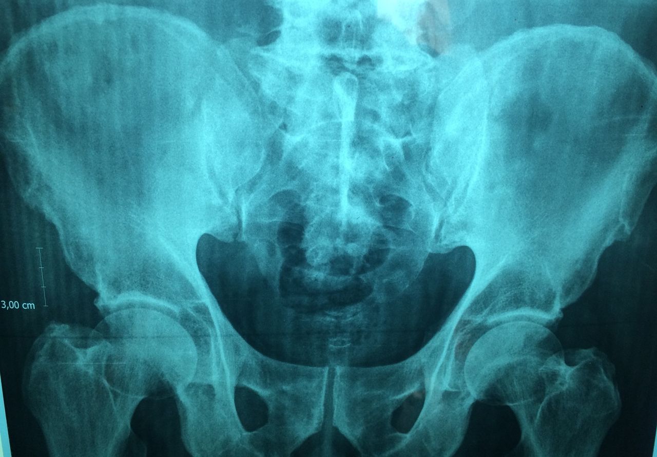
Tattooing involves introducing a minor and imperceptible modification into medical images or data entrusted to a third party. “We simply modify a few pixels on an image, for example to change the colour a little, a subtle change that makes it possible to code a message,” explains Gouenou Coatrieux. We can thus tattoo the identifier of the last person to access the data. This method does not prevent the file from being used, but if a problem occurs, it makes it very easy to identify the person who leaked it. The tattoo thus guarantees traceability. It also creates a form of dissuasion, because users are informed of this device. This technique has long been used to combat digital video piracy. Encryption and tattooing can also be combined: this is called crypto-tattooing.

Initially, LaTIM team was interested in the protection of medical images. A joint laboratory was thus created with Medecom, a Breton company specialising in this field, which produces software dedicated to radiology.

Multiple fields of application

Subsequently, LaTIM extended its field of research to the entire field of cyber-health. This work has led to the filing of several patents. A former doctoral student and engineer from the school has also founded a company, WaToo, specialising in data tagging. A Cyber Health team at LaTIM, the first in this field, has just been accredited by Inserm. This multidisciplinary team includes researchers, research engineers, doctoral students and post-docs, and includes several fields of application: protection of medical images and genetic data, and ‘big data’ in health. In particular, it works on the databases used for AI and deep learning, and on the security of treatments that use AI. “For all these subjects, we need to be in constant contact with health and genetics specialists,” stresses Gouenou Coatrieux, head of the new entity. We also take into account standards in the field such as DICOM, the international standard for medical imaging, and legal issues such as those relating to privacy rights with the application of European RGPD regulations.

The Cyber Health team recently contributed to a project called PrivGen, selected by the Labex (laboratory of excellence) CominLabs. The ongoing work which started with PrivGen aims to identify markers of certain diseases in a secure manner, by comparing the genomes of patients with those of healthy people, and to analyse some of the patients’ genomes. But the volumes of data and the computing power required to analyse them are so large that they have to be shared and taken out of their original information systems and sent to supercomputers. “This data sharing creates an additional risk of leakage or disclosure,” warns the researcher. “PrivGen’s partners are currently working to find a technical solution to secure the treatments, in particular to prevent patient identification”.

Towards the launch of a chaire (French research consortium)

An industrial chaire called Cybaile, dedicated to cybersecurity for trusted artificial intelligence in health, will also be launched next fall. LaTIM will partner with three other organizations: Thales group, Sophia Genetics and the start-up Aiintense, a specialist in neuroscience data. With the support of Inserm, and with the backing of the Regional Council of Brittany, it will focus in particular on securing the learning of AI models in health, in order to help with decision-making – screening, diagnoses, and treatment advice. “If we have a large amount of data, and therefore representations of the disease, we can use AI to detect signs of anomalies and set up decision support systems,” says Gouenou Coatrieux. “In ophthalmology, for example, we rely on a large quantity of images of the back of the eye to identify or detect pathologies and treat them better.

AI-4-Child “Chaire” research consortium: innovative tools to fight against childhood cerebral palsy

In conjunction with the GIS BeAChild, the AI-4-Child team is using artificial intelligence to analyze images related to cerebral palsy in children. This could lead to better diagnoses, innovative therapies and progress in patient rehabilitation. But also a real breakthrough in medical imaging.

The original version of this article was published on the IMT Atlantique website, in the News section.

Cerebral palsy is the leading cause of motor disability in children, affecting nearly two out of every 1,000 newborns. And it is irreversible. The AI-4-Child chaire (French research consortium), managed by IMT Atlantique and the Brest University Hospital, is dedicated to fighting this dreaded disease, using artificial intelligence and deep learning, which could eventually revolutionize the field of medical imaging.

“Cerebral palsy is the result of a brain lesion that occurs around birth,” explains François Rousseau, head of the consortium, professor at IMT Atlantique and a researcher at the Medical Information Processing Laboratory (LaTIM, INSERM unit). “There are many possible causes – prematurity or a stroke in utero, for example. This lesion, of variable importance, is not progressive. The resulting disability can be more or less severe: some children have to use a wheelchair, while others can retain a certain degree of independence.”

Created in 2020, AI-4-Child brings together engineers and physicians. The result of a call for ‘artificial intelligence’ projects from the French National Research Agency (ANR), it operates in partnership with the company Philips and the Ildys Foundation for the Disabled, and benefits from various forms of support (Brittany Region, Brest Metropolis, etc.). In total, the research program has a budget of around €1 million for a period of five years.

Chaire AI-4-Child, François Rousseau
François Rousseau, professor at IMT Atlantique and head of the AI-4-Child chaire (research consortium)

Hundreds of children being studied in Brest

AI-4-Child works closely with BeAChild*, the first French Scientific Interest Group (GIS) dedicated to pediatric rehabilitation, headed by Sylvain Brochard, professor of physical medicine and rehabilitation (MPR). Both structures are linked to the LaTIM lab (INSERM UMR 1101), housed within the Brest CHRU teaching hospital. The BeAChild team is also highly interdisciplinary, bringing together engineers, doctors, pediatricians and physiotherapists, as well as psychologists.

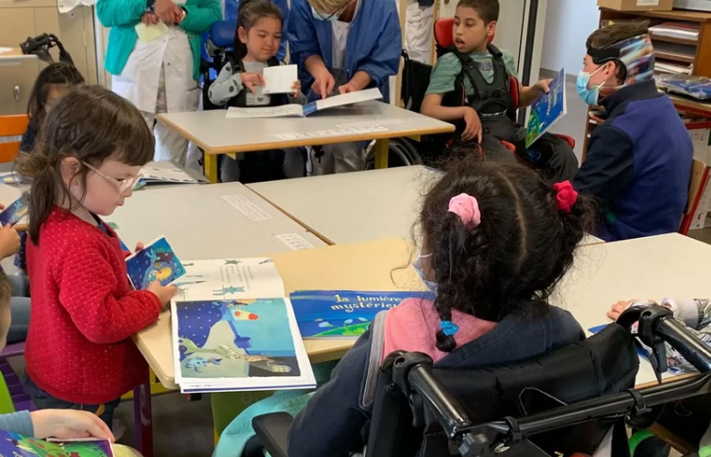
Hundreds of children from all over France and even from several European countries are being followed at the CHRU and at Ty Yann (Ildys Foundation). By bringing together all the ‘stakeholders’ – patients and families, health professionals and imaging specialists – on the same site, Brest offers a highly innovative approach, which has made it a reference center for the evaluation and treatment of cerebral palsy. This has enabled the development of new therapies to improve children’s autonomy and made it possible to design specific applications dedicated to their rehabilitation.

“In this context, the mission of the chair consists of analyzing, via artificial intelligence, the imagery and signals obtained by MRI, movement analysis or electroencephalograms,” says Rousseau. These observations can be made from the fetal stage or during the first years of a child’s life. The research team is working on images of the brain (location of the lesion, possible compensation by the other hemisphere, link with the disability observed, etc.), but also on images of the neuro-musculo-skeletal system, obtained using dynamic MRI, which help to understand what is happening inside the joints.

‘Reconstructing’ faulty images with AI

But this imaging work is complex. The main pitfall is the poor quality of the images collected, due to camera shake or artifacts during the shooting. So AI-4-Child is trying to ‘reconstruct’ them, using artificial intelligence and deep learning. “We are relying in particular on good quality views from other databases to achieve satisfactory resolution,” explains the researcher. Eventually, these methods should be able to be applied to routine images.

Significant progress has already been made. A doctoral student is studying images of the ankle obtained in dynamic MRI and ‘enriched’ by other images using AI – static images, but in very high resolution. “Despite a rather poor initial quality, we can obtain decent pictures,” notes Rousseau.  Significant differences between the shapes of the ankle bone structure were observed between patients and are being interpreted with the clinicians. The aim will then be to better understand the origin of these deformations and to propose adjustments to the treatments under consideration (surgery, toxin, etc.).

The second area of work for AI-4-Child is rehabilitation. Here again, imaging plays an important role: during rehabilitation courses, patients’ gait is filmed using infrared cameras and a system of sensors and force plates in the movement laboratory at the Brest University Hospital. The ‘walking signals’ collected in this way are then analyzed using AI. For the moment, the team is in the data acquisition phase.

Several areas of progress

The problem, however, is that a patient often does not walk in the same way during the course and when they leave the hospital. “This creates a very strong bias in the analysis,” notes Rousseau. “We must therefore check the relevance of the data collected in the hospital environment… and focus on improving the quality of life of patients, rather than the shape of their bones.”

Another difficulty is that the data sets available to the researchers are limited to a few dozen images – whereas some AI applications require several million, not to mention the fact that this data is not homogeneous, and that there are also losses. “We have therefore become accustomed to working with little data,” says Rousseau. “We have to make sure that the quality of the data is as good as possible.” Nevertheless, significant progress has already been made in rehabilitation. Some children are able to ride a bike, tie their shoes, or eat independently.

In the future, the AI-4-Child team plans to make progress in three directions: improving images of the brain, observing bones and joints, and analyzing movement itself. The team also hopes to have access to more data, thanks to a European data collection project. Rousseau is optimistic: “Thanks to data processing, we may be able to better characterize the pathology, improve diagnosis and even identify predictive factors for the disease.”

* BeAChild brings together the Brest University Hospital Centre, IMT Atlantique, the Ildys Foundation and the University of Western Brittany (UBO). Created in 2020 and formalized in 2022 (see the French press release), the GIS is the culmination of a collaboration that began some fifteen years ago on the theme of childhood disability.