ixblue

iXblue: Extreme Fiber Optics

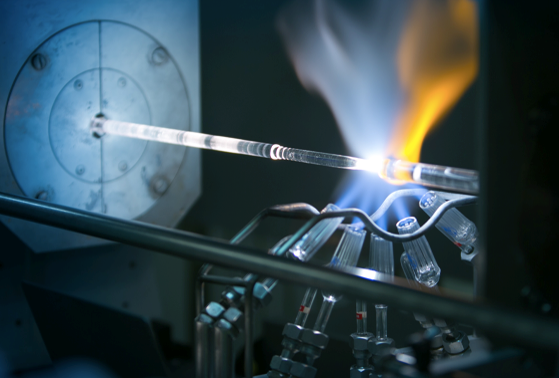
Belles histoires, Bouton, CarnotSince 2006, iXblue, a French company based in Lannion, and the Hubert Curien laboratory [1] in Saint-Étienne have partnered to develop cutting-edge fiber optics. This long partnership has established iXblue as a global reference in the use of fiber optics in harsh environments. The scientific and technological advances have enabled the company to offer solutions for the nuclear, space and health sectors. But there’s something different about these optical fibers: they’re not used for telecommunications.


Last June, iXblue and the Hubert Curien laboratory officially opened LabH6, a joint research laboratory dedicated to fiber optics. This latest development comes from a partnership that has existed since 2006 and the explosion of the internet bubble. In fact, iXblue was born from the ashes of a start-up specializing in fiber optics for telecommunications. After the disappointment experienced in the digital technology sector in the early 2000s, “we decided to make a complete U-turn, leaving telecommunications behind, while remaining in fiber optics,” explains Thierry Robin, present since the beginning and currently the company’s CTO.

A daring move, at a time when fiber optics in domestic networks was in its infancy. But it was a move that paid off. In 13 years, the young company became a pivotal stakeholder in fiber optics for harsh environments. The company owes its success to the innovations developed with the Hubert Curien laboratory. The company’s products are now used in high-temperature conditions, under nuclear irradiation and in the vacuum of space.

Measuring nuclear irradiation

One of the major achievements of this partnership has been the development of optical fibers that can measure the radiation dose in an environment. The light passing through an optical fiber is naturally diminished over the length of the fiber. This attenuation, called optical loss, increases when the fiber is under nuclear radiation. “We understand the law governing the relationship between optical loss and the radiation dose received by the fiber,” explains Sylvain Girard, a researcher at the Hubert Curien laboratory. “We can therefore have an optical fiber play the role of hundreds of dosimeters by measuring the radiation value.”

There are two advantages to this application of the fiber. First of all, the resulting data can be used to establish a continuous mapping of the radiation over the length of the fiber, whereas dosimeters provide a value from their specific location. Secondly, the optical fiber provides a real-time measurement, since the optical loss is measured live. Dosimeters, on the other hand, are usually left for days or months in their locations before the value of the accumulated radiation can be measured.

The fibers used in this type of application are unique. They must be highly sensitive to radiation in order to accurately measure the variations. Research conducted for this purpose resulted in fibers doped with phosphorus or aluminum. This type of optical fiber is currently installed in the CERN Large Hadron Collider (LHC) in Geneva during the 2-year shutdown that will continue until 2020. “This will enable CERN to assess the vulnerability of the electronic equipment to radiation and hence avoid unplanned shutdowns caused by outages,” Sylvain Girard explains.

These optical fibers are also being assessed at the TRIUMF particle accelerator center in Canada for proton therapy. This high-precision medical technique treats ocular melanomas using radiation. The radiation dose deposited on the melanoma must be very precise. “The fiber should make it possible to measure the radiation dose in real-time and stop it once the required value is reached,” the researcher explains. “Without the fiber, doctors can only determine the total dose the patient received at the end of the treatment. They must therefore accumulate three low-dose radiation sessions one after the other to come as close as possible to the total target dose.”

Surviving space

While the fibers used in dosimetry must be sensitive to radiation for measurement purposes, others must be highly resistant. This is the case for fibers used in space. Satellites are susceptible to space radiation. However, the gyroscopes satellites use to position themselves use optical fiber amplifiers. iXblue and the Hubert Curien laboratory therefore partnered together to develop hydrogen or cerium-doped optical fibers. Two patents have been filed for these fiber amplifiers, and their level of resistance has made them the reference in optical fibers for the space sector.

The same issue of resistance to radiation exists in the nuclear industry, where it is important to measure the temperature and mechanical stress in the core of nuclear reactors. “These environments are exposed to doses of a million Grays. For comparison purposes, a lethal dose for humans is 5 Grays,” Sylvain Girard explains. The optical fiber sensors must therefore be extremely resistant. Once again, the joint research conducted by iXblue and the Hubert Curien laboratory led to two patents for new fibers that meet the needs of manufacturers like Orano (formerly AREVA). These fibers will also be deployed in the fusion reactor project, ITER.

All this research will continue at the new LabH6, which will facilitate the industrial application of the research conducted by iXblue and the Hubert Curien laboratory. The stakes are high, as the uses for optical fibers beyond telecommunications continue to increase. While space and nuclear environments may seem to be niche sectors, the optical fibers developed for these applications could also be used in other contexts. “We are currently working on fibers that are resistant to high temperatures for use in autonomous cars,” says Thierry Robin. “These products are indirectly derived from developments made for radiation-resistant fibers,” he adds. After leaving the telecommunications sector and large volume production 13 years, iXblue could soon return to its origins.

[box type=”shadow” align=”” class=”” width=””]A word from the company: Why partner with an academic institute like the Hubert Curien laboratory?

We knew very early on that we wanted an open approach and exchanges with scientists. Our partnership with the Hubert Curien laboratory allowed us to progress within a virtuous relationship. In an area where competitors maintain a culture of secrecy, we inform the researchers we work with of the exact composition of the fibers. We even produce special fibers for them that are only used for the scientific purposes of testing specific compositions. We want to enable our academic partners to conduct their research by giving them all the elements they need to make advances in the field. This spirit is what has allowed us to create unique products for the space and nuclear sectors.[/box]

[1] The Hubert Curien Laboratory is a joint research unit of CNRS/Université Jean Monnet/Institut d’Optique Graduate School, where Télécom Saint-Étienne conducts much of its research.

Dominique Thers’ team at IMT Atlantique is working on XEMIS, a medical imaging device that uses liquid xenon.

Is dark matter the key to the medical scanner of the future?

A team of researchers at IMT Atlantique is developing a new type of medical scanner called XEMIS. To create the device, the team drew on their previous research in fundamental physics and the detection of dark matter, using liquid xenon technology. The first time the device was tested was using small animals. It allowed the scientists to significantly lower the injected dose, the time of the examinations, and to improve the resolution of the images produced.


This article is part of our dossier “When engineering helps improve healthcare

For the past 10 years, researchers at IMT Atlantique have been tracking dark matter as part of the XENON international collaboration. Their approach, which uses liquid xenon, currently makes them world leaders in one of the biggest mysteries of the universe: what is dark matter made of? Although the answer to this question is still waiting to be discovered, the team’s fundamental work in physics has already given rise to new ideas… in medicine! As well as detecting dark matter, the innovations produced by the XENON collaboration have proven to be extremely useful for medical imaging, as they are much more efficient than current scanners.

Improving current medical imaging techniques is one of the great challenges for medicine’s future. The personalization of patients’ healthcare and follow-up care, as well as the prediction of illnesses, mean that doctors are needing to acquire more patient data more often. However, having to remain still for 30 minutes straight is not enjoyable for patients- especially when they are now being asked to get scans more often! For hospitals, more examinations means more machines and more staff. This means that faster imaging techniques are not only more practical for patients, but also more economic for health services.

All over the world, several initiatives are competing to try and find the scanners of the future. These all have similar structures to scanners that are currently being used. “This is an area of study where investment is important” states Dominique Thers, a researcher in fundamental physics at IMT Atlantique and French coordinator of the XENON collaboration. Manufacturers are improving the scanners logically, for example, by increasing their camera size or resolution. “Since we are researching dark matter, our technology comes from another field. This means that our solution is completely different to the routes that are currently being explored”, highlights Thers, whilst reminding us that his team’s work is still in the research phase, and not for industrial use.

A xenon bath

The physicists’ solution has been named XEMIS (Xenon Medical Imaging System). Although he uses the word ‘camera’, Dominique Thers’ description of the device is completely different to anything we could have imagined. “XEMIS is a cylindrical bathtub filled with liquid xenon”. Imagine a donut led on its side and then stretched out lengthways with a hole in the middle to form a tube. The patient is led down inside the hole and surrounded by the tube’s 12cm thick wall, which is filled with liquid xenon. Although XEMIS is shaped like any other scanner, the imaging principle is completely different. In this device, the entire tube is the ‘camera’.

To understand this, let’s go into more detail. Currently, there are two main types of medical scanners: the CT scanner and the PET scanner  The CT scanner uses an X-ray source which passes through the patient’s body, and is received by a receptor on the other side of the tube. Whereas, for the PET scanner, the patient needs to be injected with a weak radioactive substance. This substance then emits ionizing radiation, which is detected by a circle of sensors that moves along the patient in the tube.

However, both devices have a size limit, which is called the parallax effect. Since the sensors face the center of the tube, their detection capacity is not the same in every direction around the patient. Therefore, image resolution is better in the center of the field of view, compared to the edge. This is why a PET scanner can only produce medical images by section, as the receptors need to be repositioned to get an accurate scan of each area of the patient.

Although XEMIS uses an injection like the PET scanner, there is no need to move sensors during the scan, as each atom of liquid xenon surrounding the patient acts as a sensor. This means that it has a large field of view that offers the same image quality in every direction. The device offers a huge advantage; now, there is no longer any need to move the sensors and work bit-by-bit. In the same amount of time as a traditional scan, XEMIS provides a more precise image; or the same quality image in a shorter amount of time.

Three photons are better than two

The highly consistent level of detection is not the only advantage of this new scanner. “The substances in traditional injections, such as those with a fluorine-18 base, emit two diametrically opposed gamma rays”, explains Dominique Thers. “When they come into contact with the xenon surrounding the patient, two things happen: light is emitted, and an electrical current is produced”. This method, which is based on contact with xenon, comes directly from the approaches used to detect dark matter.

Therefore, on both sides of the tube there are interaction points with xenon. The line between these two points passes through the patient and, more precisely, through the point where the gamma rays are emitted. Algorithms then record these signals and join them together with associated points in the patient. This is then used to build an image. “Because XEMIS technology eliminated the parallax effects, the signal-to-noise ratio of the image created by this detection method is ten times better than the images produced by classical PET scanners”, explains Dominique Thers.

As well as this, XEMIS cameras give doctors a new imaging modality called three-photon imaging! Instead of using fluorine-18, the researchers would inject the patient with scandium-44, which is made by the ARRONAX cyclotron in Nantes. This isotope emits two diametrically opposed photons, as well as a third. “Not only do we have a line that passes through the patient, but XEMIS also measures a hollow cone which contains the third proton. This passes through the emission point”. The additional geometric information allows the machine to efficiently calculate the emission point using triangulation, which ultimately leads to an even better signal-to-noise ratio.

According to Dominique Thers, “By removing the parallax effect, the device can improve the signal-to-noise ratio of a medical image of a human by a factor of ten. It can improve this ratio by another factor of ten if it uses a camera that can capture the entire patient in its field of view, and another factor of ten with the third photon. In total, this means that XEMIS can improve the signal-to-noise ratio by a factor of 1,000.” This makes it possible to make adjustments that can reduce the injected dose, or to improve the imaging protocol in order to increase the frame rate of the cameras.

Can this become an industrial scanner?

Researchers at IMT Atlantique have already demonstrated the effectiveness of XEMIS using three-photon radioactive tracers, but they have not yet tried this with living things. A second phase of the project, called XEMIS2, is now coming to an end. “Our goal now is to produce the first image of a living thing using a small animal, for example. This will demonstrate the huge difference between XEMIS and traditional scanners”, Thers explains. This is a key step in ensuring that the technology is adopted by the medical imaging community. The team are working with the Nantes University Hospital, which should help them to achieve this objective.

In the meantime, the IMT Atlantique team has already patented several innovations with Air Liquide for a xenon renewal and emptying system for the XEMIS camera. 30% of this technology uses cryogenic liquids. It is important that the team have already planned technical processes that will make it easier for health services to use XEMIS, just in case the scanner is adopted by the medical profession. This is a step forward in the international development of this unique technology.

Photographie d'un implant osseux à base de phosphate de calcium conçus par l'équipe de David Marchat.

Bone implants to stimulate bone regeneration

Mines Saint-Étienne’s Centre for Biomedical and Healthcare Engineering (CIS) seeks to improve healthcare through innovations in engineering. David Marchat, a materials researcher at CIS, is working on developing calcium phosphate-based biomaterials. Due to their ability to interact with living organisms, these bone implants can help regenerate bones.


This article is part of our dossier “When engineering helps improve healthcare

New-generation bone implants is one of David Marchat’s areas of research. The Mines Saint-Étienne chemist is developing a calcium phosphate-based biomaterial which can interact with living organisms and stimulate bone generation. Although calcium phosphate-based bone substitutes have been used in health systems for decades, their action has remained limited.

Our research focuses on the need for a bio-instructive implant, meaning one that is able to tell cells how to rebuild the bone and facilitate its vascularization.” In practice, this includes two major aspects: working on the chemical composition of calcium phosphate, and on the architecture of the implant. This is important, since existing implants are only able to regenerate small bone defects (less than 1 cm3).

When the bone is damaged too badly, it is not able to rebuild itself. A structural bone graft is then required, either from a donor (allograft), or from the patient (autograft). If the graft comes from a donor — primarily from bone banks — there is a risk that the remaining proteins cause an inflammatory response, infection or rejection. This is not the case if the graft is taken directly from another part of the patient’s body, but there is a limited quantity of harvestable bone structure. Furthermore, this involves two successive operations for the patient and the partial or complete loss of a bone.

Synthetic materials such as calcium phosphate-based biomaterials avoid these constraints. Moreover, since calcium phosphates form the mineral part of the bone, they are generally well-tolerated (i.e., non-toxic). The bone implants developed by the team meet a number of needs. The shape is personalized so as to correspond to the nature of the patient’s bone defect. This close contact between the bone margin and the implant therefore facilitates the migration of fluids, tissues and cells in the implant while also facilitating  their regeneration. The overall architecture, which ranges from the marco-scale (greater than 100 micrometers) to the nano-scale (less than 1 micrometer), is designed to “guide” this regeneration.

In addition, the composition of the calcium phosphate powder is optimized to provide chemical elements that contribute to bone formation. “We had to invent new tools and processes, in order to synthesize calcium-phosphate based powders, analyze them and then make customized bone implants,” explains David Marchat.

A new architecture

When  bone tissue is weakened a process is initiated to regenerate it, in which two types of cells act together. The first type of cells is responsible for breaking down the damaged tissue in order to recover elements that can be used by the second type of cells to rebuild the tissue. This second group of cells weave a fabric of collagen fibers – the extracellular matrix – which they then mineralize by precipitating calcium phosphate crystals. In order for the implant to effectively contribute to this bone remodeling process, the cells, blood vessels, and more generally, the new tissues, must be able to colonize it and eventually replace it.

The bone implants are modeled on several architectural levels, with pores of various sizes to promote bone regeneration and  vascular penetration. At the macro-scale level, the smallest pores  (less than 150 micrometers) confine the cells responsible for bone regeneration and stimulate their activity. Meanwhile, the largest pores (greater than 500 micrometers) allow for greater colonization by bone cells and blood vessels. “The combination of macropores of various sizes which make it possible to increase permeability and confinement,” explains David Marchat, “is essential to new bone regeneration strategies.”

To obtain the desired architecture, the researchers have developed a method based on pouring a calcium phosphate suspension — a liquid mixture containing calcium phosphate powder, water and  chemical stabilizers — into a wax mold, made using 3D printing. This “impregnated” mold is then dried and heat-treated at different temperatures in order to eliminate the wax mold and consolidate the implant.

Another question that inevitably arises is,” adds the chemist, “for a given application, how long does the structure have to remain in the body so that the bone has time to regenerate itself,” adds the chemist. If the implant deteriorates too slowly, it will block bone formation. But if, on the other hand, it deteriorates too quickly, it will not be able to serve as a scaffold for bone formation. “It’s hard to estimate the right balance between the two.

An ambitious engineering project for the testing phase

There are currently two ways to carry out biological assessments of these bone implants, which are used at different stages of testing. Standard in vitro cultures have the advantage of allowing for direct observation through a microscope but do not reproduce real conditions inside the body. In vivo experimentation with implantation inside an animal recreates these conditions more closely and provides what is referred to as a “physiological” environment, although animal and human physiology are different, and it is difficult to access the information. These testing stages are crucial, but researchers would like to move away from animal testing.

This is precisely the aim of an ambitious project: developing a 3D bioreactor with human cells to mimic human physiology. Such a structure would provide conditions equivalent to the human body and allow for direct observations, thereby making animal testing unnecessary for majority of validation phases for medical devices or medications. This project calls for expertise in fluid mechanics and a greater understanding of the human body and its workings. Other research topics in healthcare medical engineering have a similar aim. One such example are Organ-On-Chips, microfluidic chips that act as artificial cell cultures to simulate the inner workings and physiological activity of human organs.

nose

An “electronic nose” analyzes people’s breath to help sniff out diseases

In partnership with IMT Atlantique, a team of researchers at IMT Lille Douai have developed a device which can measure the level of ammonia in someone’s breath. The aim of the artificial nose is to use this device to create a personalized follow-up care for patients affected by chronic kidney disease.  Eventually, the machine could even allow doctors to detect the disease in undiagnosed people.


This article is part of our dossier “When engineering helps improve healthcare

In the human body, the kidneys’ main role is to remove toxins which are carried in the blood. However, when a person suffers from chronic kidney disease, this filtration function no longer works to the same standard. In France, the disease affects around 5.7 million people and can range from causing a degree of impairment, to terminal. Around 76,000 people are terminally affected in France, which means that their kidneys cannot filter their blood at all.

For these patients, the only options are to either wait for a transplant or face extensive treatment which hugely affects their daily life. It is therefore essential for doctors to be able to detect this silent and progressive disease early enough to slow down the effects. At the moment, doctors use blood or urine tests to identify the disease. But, to make it easier to diagnose, scientists are exploring another route: breath analysis. Studying the substances in the air we breathe out can provide valuable information about a person’s health.

Ammonia: a key element

In order to make this possible, two teams of researchers from IMT Lille Douai and IMT Atlantique have been working in partnership with the nephrology department at the Lille University Hospital. For the past three years, they have been developing a compact and turnkey device for doctors. The device is an ‘electronic nose’, a system made up of several sensors that can measure the specific concentration of a substance in someone’s breath.

In the case of chronic kidney disease, the substance being measured is ammonia. Ammonia is mainly produced by intestinal bacteria and is supposed to be filtered from the body by the kidneys. Previous studies have established a concentration threshold for levels of ammonia in a person’s breath, which doctors can then use to determine the likelihood that a patient has chronic kidney disease.

Ammonia offers some resistance

But how can you measure this compound using a portable device? The scientists used a series of sensors which react in the presence of ammonia, as Caroline Duc, team member and researcher at IMT Lille Douai, explains. “The sensors are made from two electrodes with a sensitive surface placed on top of them. The resistance of this surface varies depending on the amount of ammonia present”. When used, the device’s ability to resist electrical current increases when ammonia is present and returns to its initial state when the ammonia disappears. This therefore allows scientists to measure the level of the molecule in a patient’s breath.

Additionally, each sensor in the artificial nose has a unique composition. As Caroline Duc points out, “it is very complicated to use a material whose resistance varies when one type of gas is present”. This is why scientists decided to increase the accuracy of the analysis by combining several different sensors which have different responses to ammonia.

During the tests, the electronic nose was periodically exposed to a person’s breath. This resulted in an increase in the resistance when ammonia was present, and a decrease when the sensors were no longer exposed to the exhaled air.  Several factors were then measured and analyzed using statistical processing algorithms.

These algorithms rely on tools such as machine learning. The only difference with this case was that here, these tools were applied using small amounts of data and  supervised learning to categorize the different types of breath. In other words, the algorithms were taught using a dataset that had already classified breath which belongs to a healthy, ill or ‘uncertain’ individual. New profiles were then passed through this algorithm, so that they could be classed into these three categories.

Breath: a complex compound

To date, the first prototype developed is around 15cm in length and contains between 10 and 13 sensors.  The device was tested in a laboratory using artificial breath, which allowed the teams to verify that it could distinguish a healthy individual from someone who was ill, using the criteria defined in up-to-date scientific literature.  Then, experiments were carried out in clinics with patients suffering from chronic kidney disease. The idea was to measure the concentration of ammonia in their breath, before and after dialysis. The results demonstrated that there was a reduction in ammonia after the treatment.

However, they also highlighted the limitations of using a single marker. Before dialysis, some patients had levels of ammonia that were similar to those of a healthy person. Measuring the amount of ammonia in a person’s breath did not give a reliable diagnosis for chronic kidney disease.  This has caused scientists to launch a new study in order to identify other biomarkers characteristic of the disease.

More generally, this reflects the difficulty of conducting a reliable breath analysis, which can include both the air we breathe in as well as out. “Initially, I think clinical breath analysis will be developed for personalized patient follow-up care, rather than for diagnosis”, says Caroline Duc. For example, by measuring how a patient’s ammonia levels change in response to medication, doctors will be able to monitor the effectiveness of a treatment and then adapt it based on the results.

What is the future of follow-up care for other illnesses?

Researchers at IMT Lille Douai will continue to work on improving the electronic nose. At the moment, patients’ breath is collected and sealed in an airtight bag and is then analyzed in a laboratory. Consequently, the team’s aim is to develop a functional and completely autonomous prototype which would give doctors real-time results. However, this raises several new issues, such as the study of fluids, controlling the speed that the air is exhaled, etc. As well as this, to improve their data analysis, Caroline Duc and her colleagues have started a partnership with researchers who specialize in data handling at Télécom SudParis.

Moreover, the team is involved in a European-wide project which aims to identify biomarkers for lung cancer, and then create a multi-sensor system that is specifically designed to detect these substances. IMT Lille Douai’s expertise will be especially useful for this second objective.

This electronic nose, capable of sniffing out illnesses such as chronic kidney disease, is therefore still in the early stages of development, and needs a lot of work before it can really be used by doctors and their patients. But doctors are waiting with bated breath; in several years’ time it could be a breathtaking medical innovation!

Article written (in French) for I’MTech by Bastien Conteras

ALGIMEL is an environmentally-friendly material which is used in a wide range of projects.

ALGIMEL, a ‘marine’ polystyrene

In the future, materials will not only need to be more efficient; it will also be essential that they are environmentally friendly. With this in mind, researchers from IMT Mines Alès who specialize in bio-sourced materials are working on this project.  Over the past few decades, they have been trying to develop environmentally-friendly alternatives to the most polluting materials. One of their latest designs is a naturally occurring polymer foam which can replace several of polystyrene’s uses. Eric Guibal and Thierry Vincent tell us about their work.


What is the material you have developed like?

Eric Guibal: We see it as a material with similar properties to polystyrene. It is a low-density foam with a structure that is mainly made up of biopolymers. This material, named ALGIMEL, is mostly made up of alginate, a natural polymer which is found in the cell wall of brown algae. The team’s expertise is mainly focused on the synthesis and composition of these foams.

Why did you choose this type of biopolymer?

Thierry Vincent: From the beginning, our aim was to develop the most environmentally-friendly material possible. This decision goes far beyond the choice of polymer as during the synthesis of the material we don’t use any toxic products. For example, when we are developing the foam, we don’t use any chemical solvents.  We don’t use products which could be dangerous for the technicians. The additives which we use to improve the properties of the material are also natural. Our manufacturing processes have low energy consumption and the drying process is carried out at a maximum temperature of 50°C. Some synthetic products are still used during manufacturing, but they make up less than 1% of all the materials that we use, and we are working on replacing them with bio-sourced materials.

Why did you want to develop these foams?

EG: Our aim was to produce an insulating material which was also an alternative to polystyrene. This is because polystyrene takes several hundred years to biodegrade, pollutes water and releases several toxic substances when it burns. Although our biopolymer foam has similar thermal insulating properties to polystyrene, it is also different due to its outstanding fire-resistant properties. ALGIMEL also has a controlled lifespan. When it is being used, the material is stable; however, at the end of its life, its biodegradable properties mean that it can be put in the household compost.

What could this material be used for?

TV: This material is light and extremely versatile, which means it can have many uses. As well as its thermal insulation and flame-retardant properties, its surface properties can be modified by applying biopolymers which make the foam hydrophobic, so it can be used in humid environments. To increase its mechanical strength, you can easily add plant-based fibers or mineral fillers. We can also add pigments or dyes, both on the interior and the surface, to change its appearance. If it’s used with other materials such as wood, fabrics, leather, etc., either in its composite form or as a sandwich compound, the foam has many uses. These include decoration, packaging, bags, clips fashion and luxury items, etc.

How did your research lead you to this material?

TV: For around thirty years, our work has focused on developing biopolymers. Our main area of experience is with alginate, as well as other biopolymers such as chitin and chitosan, which are made from crustacean shells. We have always directed our research towards developing materials that are more environmentally friendly. ALGIMEL is the result of all the skills we have acquired during our research.

Will the material soon be used outside of your laboratory?

EG: We are currently working with organizations that specialize in technology transfer, including SATT AxLR Occitanie Est. We are also lucky enough to have a contract with Institut Carnot M.I.N.E.S and the Carnot Carats network. Today, we are in the process of raising process quality and pre-industrialization. In collaboration with several partners, we are working on improving the design of our foams so they can be used in decorative, fashion and luxury products. We know that the development of this type of material is in line with many announcements made by the public authorities and industrialists. The most recent example is Emmanuel Macron’s aim of making the fashion and luxury goods industries more sustainable, which he entrusted to François-Henri Pinault. All of this shows that this product has a promising future ahead of it.


microorganisms

When microorganisms attack or repair materials

Some microorganisms can seriously damage structures made of concrete or stone, leading to billions of euros in damage. Others, on the contrary, have a positive effect as they are able to heal micro-cracks.


They are microscopic, but can cause billions of euros in damage. Microorganisms, such as bacteria, fungi and algae, are ubiquitous in the environment and can develop under some conditions on the surface of buildings and structures. Their impacts should not be neglected. Facades, water treatment plants and sewer pipes must all be monitored, repaired and better designed to withstand these attacks.

Costly deterioration

Microorganisms form biofilms that adhere to the surface of materials and interact with them. In certain cases, they deteriorate the materials,” explains Christine Lors, professor at the Center for Educating, Research and Innovation (CERI) in Materials and Processes at IMT Lille Douai. “This causes several billions of euros of damage, particularly for sewage systems.” Indeed, most of the large-diameter pipes that carry our wastewater to treatment plants are made of concrete, and their premature deterioration is very costly. Microorganisms develop on the surface of concrete as well as on the stones of buildings, such as churches. This does not always endanger the monuments but can generate significant costs in cleaning the biological fouling, as in the case of facings. They can also affect health in the indoor environments of buildings, through the proliferation of fungi, which can lead to pathologies for occupants.

In general, microorganisms do not directly deteriorate the structures. They produce molecules through their metabolism that alter the materials. For example, sulfur-oxidizing bacteria derive their energy from sulfur compounds such as hydrogen sulfide with its rotten-egg odor present in wastewater. They transform it into sulfuric acid, which is very aggressive for concrete. Nitrifying bacteria found in wastewater treatment plants, on the other hand, produce nitric acid from nitrogen pollution. Finally, in the biomethanation plants, which operate in the absence of oxygen, the acidogenic bacteria produce organic acids, such as acetic, propionic and butyric acids.

Solutions exist

How to prevent or at least limit biodeterioration? First of all, by working on the surface of the materials to reduce the adhesion of the biofilm. The rougher the surface, the easier it is for microorganisms to adhere to it, finding in each roughness an anchor point. The material’s surface tension also plays a role in the adhesion of the biofilm.

The other means of fighting biodeterioration is changing the material itself. Some cements resist better because they neutralize the acid produced without rapidly decreasing their own pH, thus preventing, as in the case of sewage systems, the development of the most acidophilic species. Another important factor to consider is that unaltered concrete is highly basic and very few microorganisms can develop on it. A concrete designed so that its surface pH decreases weakly limits the colonization of its surface.

Finally, for existing structures that tend to deteriorate, it is possible to add a layer of high-strength fiber-reinforced concrete that protects the underlying initial concrete. This addition is costly, but not as expensive as replacing the entire material for large-diameter pipes and sewage collectors.

Useful microorganisms

So far, this depiction of microorganisms has painted them as a scourge to be systematically eradicated from all structures. However, in certain cases, they are actually beneficial. “Some bacteria have a positive effect, by depositing calcium carbonate (limestone) on the material’s surface, they protect them and fill in micro-cracks,” Christine Lors explains. “This is the case for bacteria of the Bacillus genus, which, when breathing, produces carbon dioxide, which combines with calcium to form calcium carbonate.” This phenomenon is of great interest to EDF, which collaborates with the Materials and Processes CERI on the issue of repairing micro-cracks in nuclear enclosures. The goal: to develop a bioprecipitation process to repair these micro-cracks, especially in inaccessible areas.

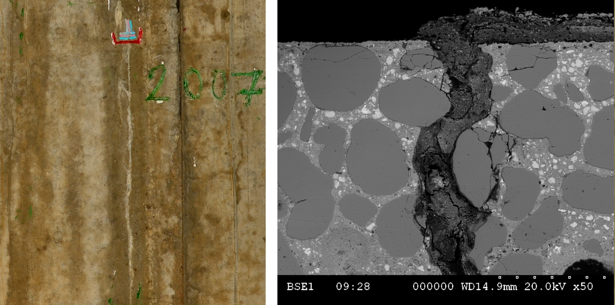
Microorganisms can heal cracks in a wall. The bioprecipitated white calcium carbonate that seals the crack appearing as a white line (left side). A scanning electron microscopy image (right side) shows the filling phenomenon.


We began by developing laboratory tests to understand the bioprecipitation mechanisms”, the researcher explains. “This required understanding under which conditions the calcium carbonate producing bacteria adhere to the micro-cracked material and then fills in the micro-crack. Then, we have developed a process to control the growth of these bacteria by optimizing their potential to produce calcium carbonate according to many parameters (temperature, nutrients, oxygenation, etc.). Finally, we conducted tests on a model from EDF that reproduces at 1/3 scale a nuclear reactor enclosure. The initial tests are promising: the micro-cracks are clogged and resist the application of a pressure of 5 bars corresponding to the decennial pressurization tests.”

Bioprecipitation process to repair nuclear enclosures

The current bacterial strain capable of healing micro-cracks comes from a laboratory collection. In the future, the objective is to use indigenous strains taken from the nuclear site, previously identified, and whose potential for production of calcium carbonate is verified. These indigenous strains are indeed better adapted to the environmental conditions of the site. When will this technique be applicable under real conditions in nuclear enclosures? “We are currently checking the feasibility, through a PhD thesis underway on this topic that is co-funded with EDF,” Christine Lors explains. “This method could be operational by the end of the PhD thesis.” The Materials and Processes CERI is also planning to create a start-up to develop this process.

Regardless of whether their effects are negative or positive, interactions between microorganisms and civil engineering materials must be taken into account more effectively. This is a standardization issue. “The current standards for structures incorporate both mechanical and chemical aspects, but the impact of microorganisms is by no means taken into account,” Christine Lors explains. “This should be done at the European level, with standardized tests. This is one of the conditions required in order for manufacturers to test their products and conduct research to improve them by taking into account biological alteration.” Thus, there is a big wait for standardization organizations.

Article written (in French) by Cécile Michaut, for I’MTech

Also read on I’MTech

Polybioskin

Polybioskin, natural skin through more ethical products

Projets européens H2020Skin contact products, whether for medical, sanitary or cosmetic purposes, have two major drawbacks: they are neither recyclable nor biodegradable. The Polybioskin H2020 project aims to correct these aspects which are out of step with consumers’ growing environmental awareness and concerns. Launched two years ago, the Polybioskin project brings together 12 European partners, including IMT Mines Alès, and will come to a close in May 2020. José-Marie Lopez-Cuesta, a materials researcher at IMT Mines Alès, presents the challenges involved in this project.


Could you describe the context of the Polybioskin project?

José-Marie Lopez-Cuesta: Skin is the human body’s most important organ and our first line of defense against external agents. Cosmetics, along with skin care and biomedical products, are developed to allow for direct contact with the skin or to protect it. These products represent a significant market which includes both low-cost and high-performance products. Today, most of these products are obtained from polymers based on fossil-fuel resources which are neither recyclable nor biodegradable.

So what is the aim of this project?

JMLC: Polybioskin must enable the industrial development of bio-based, renewable solutions for antimicrobial, antioxidant and absorbent applications for skin contact products. The three target markets are sanitary, cosmetic and biomedical products.

What scientific problems must you respond to?

JMLC: The products developed must be economically competitive and have a renewable content of 90%. We also strive to reduce the environmental footprint, through different end-of-life scenarios for the products developed. Life-cycle analyses must demonstrate their sustainable character and their compliance with safety regulations.

Who are your partners for this project and how is their collaboration important to your work?  

JMLC: The Polybioskin consortium combines the expertise of 12 partners from 7 European countries. We already have relationships with several of the academic partners through the ENMAT research network (European Network on Materials). And there are also non-academic partners who play an important role. The industry partners included in the BBI association are stakeholders in the definition of calls for projects aiming to promote bioplastics. This project can also help launch new collaborations as part of PhD theses and prepare responses to new calls for H2020 projects.

Polybioskin draws on expertise in biology, chemistry, material sciences, nanotechnologies and other fields. How have researchers from IMT Mines Alès contributed to the project?

JMLC: IMT Mines Alès develops superabsorbent structures for diapers and polymer films based on alloys formulated for beauty mask applications. The goal is to develop these structures using only bio-based components through chemical modifications and plastics processes. Different sources of cellulose have been used to synthesize absorbent structures. IMT Mines Alès also contributes to analyzing the life-cycle of all the components developed over the course of the project. To do so, we use specific tools including databases on the energy consumption and impact of the different components.

What are the current and future steps for Polybioskin ?

JMLC: The project is two-thirds complete. We’re currently in the pilot phases in order to develop prototypes. These prototypes are developed by assembling the materials developed in the earlier stages of the project. Publications and scientific papers have already been produced. In addition, a consortium agreement has been signed with the industry partners involved in the project. This will help us manage the technology transfer of the results at the end of the project, so that our scientific results can directly contribute to bringing innovative products to the market.

[divider style=”solid” top=”20″ bottom=”20″]

Polybioskin project partners

[divider style=”solid” top=”20″ bottom=”20″]


Romain Quey

Romain Quey, all about polycrystals

A recent winner of a CNRS Bronze medal, Romain Quey is a materials science researcher at the Georges Friedel[1] laboratory at Mines Saint-Étienne. He focuses especially on the deformation of metallic polycrystalline materials. In the following interview for I’MTech, he presents his research, between synchrotron radiation and digital simulation.  


Could you define what a polycrystalline material is?

Romain Quey: A lot of the materials around us are generally polycrystalline:  metals, which are my primary focus, as well as rocks, ice, certain polymers, etc. If you observe the structure of such a material, for example with an optical or electronic microscope, you’ll see that it is composed of a large number of grains which vary in size between approximately 10 and 100 micrometers in diameter. The grains have a crystalline structure, meaning that their atoms are arranged in a regular pattern. This arrangement can be cubic, hexagonal etc. In the simplest polycrystalline materials, all the grains have the same crystalline structure, but the orientation of these crystalline structures varies from one grain to another. Since a crystalline grain does not deform in the same way in different directions of space, this creates a certain disorder. That’s the basic principle!

Why is this microscopic structure important in the behavior of polycrystalline materials?  

RQ: Take aluminum alloys or steels, for example, which are everyday materials. At your level, you may have the impression that a material like this deforms in a uniform manner. However, on the microscopic level, it’s a different story: different grains tend to deform in different ways, and the resulting internal stresses are incredibly heterogeneous. It’s these microscopic aspects that govern the macroscopic mechanical properties of the materials, like how the material breaks, or how its polycrystalline structure evolves during deformation.

Your research mainly involves studying the evolution of these microstructures when the material is deformed. How do you go about this?

RQ: There are two main parts to this research: experiments to observe them, and digital simulation, to try to reproduce them. For the experimental part, I increasingly use synchrotron X-ray radiation.   We use a synchrotron to obtain intense radiation, with a high flux of X-rays, which can easily pass through several millimeters of a material. Using synchrotron radiation, we can observe what happens within a sample during its deformation in a 3D, non-destructive manner. We can obtain 3D images of the initial or deformed material with close to a 1-micrometer resolution. These methods have mainly been developed and implemented at ESRF, in Grenoble, but we have contributed as well.

Is this where simulation comes into play in your work?

RQ: Yes, since once we have observed the material and its evolution during deformation, we want to try to reproduce it by simulation, using the information we’ve put into the models. These models all integrate the same basic mechanisms of deformation, but there are different types of models and all of them involve parameters which are not always well known. We try to obtain a simulation result that’s as close to the experimental observation as possible. This is the aim of the ANR 3DPLASTICITY project currently being carried out. In addition to validating models, this helps us develop a greater understanding about how materials behave and evolve.

This research focusing on simulating the behavior of polycrystalline materials led to your creating a software: Neper. What is its purpose?

RQ: When researchers or engineers want to make a simulation-based calculation for a polycrystalline material, they must first create a virtual polycrystal. Neper makes it possible to do this. It allows us to generate a polycrystalline microstructure and then to mesh it with finite elements. The microstructure can be generated from experimental data available to the user: an image, distribution of grain sizes, crystalline orientations, etc. It’s a powerful but easy-to-use software, which responds to real needs and can be applied to a number of fields.  Applications mainly involve metals but also include rocks, ice or polymers, and can be used for research in mechanics as well as in thermal engineering, magnetism and acoustics. The software is therefore regularly used by researchers and industry players in a wide variety of disciplines.

Beyond its academic contribution, what are the applications of your research for engineering?

RQ: In parallel with the previously-mentioned complex models, which can only be used for computing clusters, we also develop “simplified” models of the evolution of microstructures during deformation, which integrate only a few key mechanisms. These models are very rapid and can be integrated in simulation chains for shaping processes already in place in industry, which aim to predict the microstructures produced by thermomechanical treatments. For industry, it is crucial to control the microstructures of materials to optimize their properties.

What are your current research perspectives for the topic of polycrystalline materials?

RQ : We’re at the point where our 3D experiments allow us to study the deformation of polycrystalline microstructures accurately. But the digital models still have to be validated. The challenge is now to compare the experiments and simulations more closely, in order to draw conclusions about the validity of the models, and about what that means in terms of the materials’ behavior and potential ways to improve their use properties. We’re getting to a point where we’ll be able to respond to a lot of uncertainties. We’re also studying the behavior of materials with new and increasingly complex microstructures and strive to obtain even more accurate results. Experimenting, modeling microstructures and simulating their behavior are still essential aspects of this work.

[1]  Georges Friedel Laboratory: joint research unit between CNRS/Mines Saint-Étienne.


carbon fibre

Recycling carbon fibre composites: a difficult task

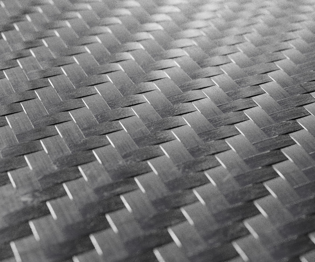
Carbon fibre composite materials are increasingly widespread, and their use continues to rise every year. Recycling these materials remains difficult, but is nevertheless necessary at the European level for environmental, economic and legislative reasons. At IMT Mines Albi, researchers are working on a new method: vapo-thermolysis. While this process offers promising results, there are many steps to be taken before a recycling system can be developed. 


The new shining stars of aviation giants Airbus and Boeing, the A350 and the 787 Dreamliner, are also symbols of the growing prevalence of composite materials in our environment. Aircraft, along with wind turbines, cars and sports equipment, increasingly contain these materials. Carbon fibre composites still represent a minority of the composites on the market — far behind fiberglass — but are increasing by 10 to 15% per year. Manufacturers must now address the question of what will become of these materials when they reach the end of their lives? In today’s society, where considering the environmental impact of product is no longer optional, the recycling issue question cannot be ignored.

At IMT Mines Albi, scientific research being carried out by Yannick Soudais[1] and Gérard Bernhart[2] addresses this issue. The researchers in polymer and materials chemistry are developing a new process for recycling carbon fibre composites. This is no small task, since it requires separating the fibre present in the form of a textile or unidirectional filaments from the solid resin polymer that forms the matrix in which it is plunged.  Two main processes currently exist to try to separate the fiber from the resin: pyrolysis and solvolysis. The first consists of burning the matrix in an inert nitrogen atmosphere in order to avoid burning part of the fiber. The second is a chemical method based on solvents, which is very laborious, because it requires high temperature and pressure.

The process developed by the Albi-based researchers is called  “vapo-thermolysis” and combines these two processes. At present, it is one of the most promising solutions in the world to move toward the wide-scale reuse of carbon fibres. Besides Albi, only a handful of other research centers in the world are working on this topic (mainly in Japan, China and South Korea). “We use superheated water vapor which acts as a solvent and induces chemical degradation reactions,” explains Yannick Soudais.  Unlike with pyrolysis, there is no need for nitrogen. And compared to the traditional chemical method, the process takes place under atmospheric pressure. In short: vapo-thermolysis is easier to implement and master on an industrial scale.

After recovery, reuse

The simplest way to reuse carbon fibres is to spread out the bundle of interlinked fibres on a flat surface and reuse it in this form, as a carpet. They will therefore be used to make composites for decorative parts rather than structural parts. The size of the recovered fibres can also be further reduced to be used as reinforcements for polymer pellets. This approach makes it possible to produce automobile parts using injection, for example. Demonstrations illustrating this type of reuse have been carried out by the researchers in collaboration with the Toulouse-based company Alpha Recyclage Composites (ARC).

But the real challenge remains being able to reuse these fibers for higher-performance uses.  To do so, “we have to be able to make spun fibers from short fibres,” says Gérard Bernhart. “We’re carrying out extensive research on this topic in partnership with ARC because so far, no one in the world has been able to do that.” These prospects involve techniques specific to the textile industry, which is why the researchers have formed a partnership with the French Institute of Textiles and Clothing (IFTH). For now, the work is only in its exploratory stages and focuses on determining technologies which could be used to develop reshaping processes. One idea, for example, is to use ribbed rollers to form homogenous yarns, then a card to create a uniform voile, followed by a drawing and spinning stage.

For manufacturers of composite parts, these prospects open the door to more economically-competitive materials. Of course, recycling is an environmental issue and certain regulations establish standards of behavior for manufacturers. This is the case, for example, for automobile manufacturers, who must ensure, regardless of the parts used in their cars, that 85% of the vehicle mass can be recycled when it reaches the end of its life. But mature, efficient recycling processes also help lower the cost of manufacturing carbon fibre composite parts.

When the fibre is new it costs €25 per kilo, or even €80 per kilo for fibres produced for high-performance materials. “The price is mainly explained by the material and energy costs involved in fibre manufacturing,” says Gérard Bernhart. Recycled fibres would therefore lead to new industrial opportunities. Far from being unrelated to the environmental perspective, this economic aspect could, on the contrary, be a driving force for developing an effective system for recycling carbon fibres.


[1] Yannick Soudais is a researcher at the Rapsodee laboratory, a joint research unit between IMT Mines Albi/CNRS
[2] Gérard Bernhart is a researcher at the Clément Ader Institute, a joint research unit between IMT Mines Albi/ISAE/INSA Toulouse/University Toulouse III-Paul Sabatier/CNRS


catenary catenaries

A mini revolution in railway catenaries

The decade-long ACCUM project carried out by SNCF, Stratiforme Industries, the Valenciennes Railway Testing Center and IMT Lille Douai has led to the development of a new catenary cantilever system for railways. This advance represents a major change in this field, where equipment has seen little change over the past 50 years.


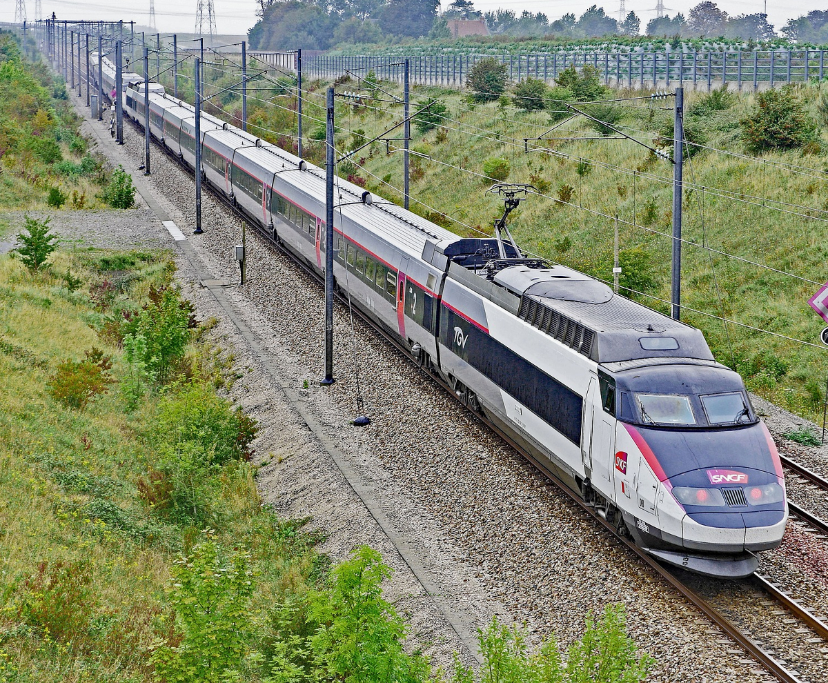
When asked to draw a train on a railway track, odds are that most people would not think to include the poles along the railway. Yet these vertical support structures, placed every 20 to 60 meters along electrified railway lines, are essential for supporting the overhead lines that power the trains. The portion located at their peak, which is responsible for holding the wires, plays an especially crucial role. It is composed of a cantilever system, which must at once support the contact wire at a constant height with centimeter precision (for high-speed lines), and withstand significant mechanical constraints arising from the voltages applied to the wires, while ensuring the electric insulation between the wire and the pole.

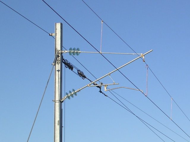
A traditional catenary cantilever is made up of numerous parts which take a long time to assemble.


The catenary cantilever system is an extremely sensitive piece of equipment which has remained virtually unchanged for a half a century. Composed of some hundred parts assembled to form a triangular metal tube, current cantilevers are a real puzzle to assemble and adjust. “Since the stresses are triangulated on the structure, when an adjustment is made at one location within the system, everything is shifted, and it all has to be adjusted again,” explains Patrice Hulot. An engineer at Lille Douai, he contributes to the ACCUM[1] project, which aims to simplify catenary cantilevers.

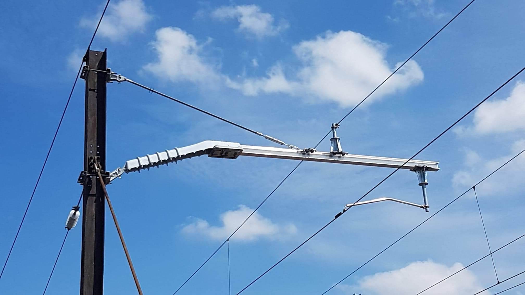
Les armements caténaires ACCUM sont en composites, et bien plus simples à monter pour les opérateurs.

The ACCUM catenary cantilevers are made from composites, and are much easier for operators to assemble.


This modernization project has been carried out over the last ten years by the SNCF and Stratiforme, a company that specializes in composite materials. In 2019, it culminated in the installation of 50 prototypes on test lines at the Railway Testing Centre (CEF), followed by installations on commercial lines. It represents a revolution for SNCF lines, and offers catenary operators the first in-depth modification of this system in fifty years.

A universal catenary cantilever system

What sets apart the new cantilevers developed through the ACCUM project is that they are composed of a limited number of parts to assemble on site. “Everything is delivered 80% pre-assembled,” says David Cnockaert, project manager at Stratiforme. “And these final components to be assembled make it possible to cover all the different types of post configurations and railways.” Furthermore, these new fittings can be used for 1,500 volt and 25,000 volt lines alike. The flexibility of this system allows it to be described as universal, since it can be adapted to all types of electrification, hence its name –ACCUM, the French acronym for “universal multi-voltage composite catenary cantilever.”

The first systems to be installed demonstrated how easy they are to assemble compared to the previous systems, with the major benefit being the time needed for adjustment and fine-tuning, which represents up to 50% of the total time needed to install or replace cantilevers. This therefore makes it possible to reduce the time required to set up the cantilevers,  significantly increasing the availability of railways during renovation work while reducing installation costs and the duration of work to put in new lines. These results are even more satisfactory since they are only the initial results. “Operators had decades of experience to optimize the installation of the old systems, so the installation of the new cantilevers will clearly take less time in the months and years to come,” says Patrice Hulot.

This innovation has received praise within the industry. The project was rewarded at JEC World in March — the global composite materials show — by an Innovation Award. It should be noted that while, for the moment, the new catenary cantilever has only been implemented on the French railways, this mini revolution in railway equipment has the potential for international success. The SNCF is a global leader in the high speed rail sector, in terms of both rolling stock and infrastructure. Its competitors therefore closely monitor developments in the field. This means that Japan or North Africa could soon be added to the list of future markets for the universal composite catenary cantilever.


[1] FUI ACCUM project, co-funded by BPI France and the Hauts-de-France region and accredited by the i-TRANS competitiveness cluster. Project leader: Stratiforme. Partners: IMT Lille Douai, ARMINES, Railway Testing Center (CEF) and the SNCF Network.