aneurysm

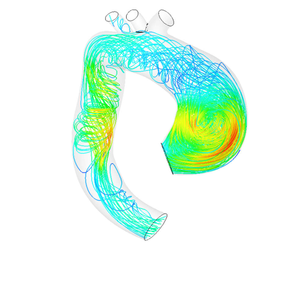
A digital twin of the aorta to prevent aneurysm rupture

15,000 Europeans die each year from rupture of an aneurysm in the aorta. Stéphane Avril and his team at Mines Saint-Étienne are working to better prevent this. To do so, they develop a digital twin of the artery of a patient with an aneurysm. This 3D model makes it possible to simulate the evolution of an aneurysm over time, and better predict the effect of a surgically-implanted prosthesis. Stéphane Avril talks to us about this biomechanics research project and reviews the causes for this pathology along with the current state of knowledge on aneurysms.


Your research focuses on the pathologies of the aorta and aneurysm rupture in particular. Could you explain how this occurs?   
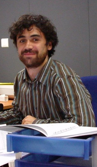
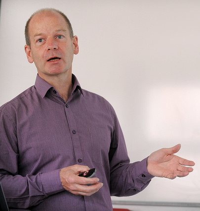
Stéphane Avril

Stéphane Avril: The aorta is the largest artery in our body. It leaves the heart and distributes blood to the arms and brain, goes back down to supply blood to the intestines and then divides in two to supply blood to the legs. The wall of the aorta is a little bit like our skin. It is composed of practically the same proteins and the tissues are very similar. It therefore becomes looser as we age. This phenomenon may be accelerated by other factors such as tobacco or alcohol. It is an irreversible process that results in an enlarged diameter of the artery. When there is significant dilation, it is called an aneurysm. This is the most common pathology of the aorta. The aneurysm can rupture, which is often lethal for the individual. In Europe, some 15,000 people die each year from a ruptured aneurysm.

Can the appearance of an aneurysm be predicted?

SA: No, it’s very difficult to predict where and when an aneurysm will appear. Certain factors are morphological. For example, some aneurysms result from the malformation of an aortic valve: 1 % of the population has only two of the three leaflets that make up this part of the heart. As a result, the blood is pumped irregularly, which leads to a microinjury on the wall of the aorta, making it more prone to damage. One out of two individuals with this malformation develops an aneurysm, usually between the ages of 40 and 60. There are also genetic factors that lead to aneurysms earlier in life, between the ages 20 and 40. Then there are the effects of ageing, which make populations over 60 more likely to develop this pathology. It is complicated to determine which factors predominate in relation to one another. Especially since if at 30 or 40 an individual is declared healthy and then starts smoking, which will affect the evolution of the aorta.

If aneurysms cannot be predicted, can they be treated?

SA: In biology, extensive basic research has been conducted on the aortic system. This has allowed us to understand a lot about what causes aneurysms and how they evolve. Although specialists cannot predict an aneurysm’s appearance, they can say why the pathology appeared in a certain location instead of another, for example. For patients who already have an aneurysm, this also means that we know how to identify the risks related to the evolution of the pathology. However, no medication exists yet. Current solutions rely rather on surgery to implant a prosthesis or an endoprosthesis — a stent covered with fabric — to limit pressure on the damaged wall of the artery. Our work carried out with the Sainbiose joint research unit [run by INSERM, Mines Saint-Étienne and Université Jean Monnet], focused on gathering everything that is known so far about the aorta and aneurysms in order to propose digital models.

What is the purpose of these digital models?

SA: The model should be seen as a 3D digital twin of the patient’s aorta. We can perform calculations on it. For example, we study how the artery evolves naturally, whether or not there is a high risk of aneurysm rupture, and if so, where exactly in the aorta. The model can also be used to analyze the effect of a prosthesis on the aneurysm. We can determine whether or not surgery will really be effective and help the surgeon choose the best type of prosthesis. This use of the model to assist with surgery led to the creation of a startup, Predisurge, in May 2017. Practitioners are already using it to predict the effect of an operation and calculate the risks.

Read on IMTech: Biomechanics serving healthcare

How do you go about building this twin of the aorta?  

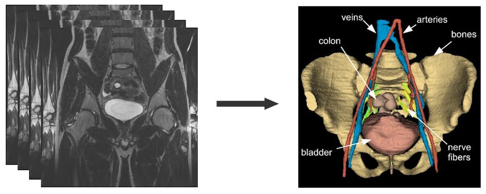
SA: The first data we use comes from imaging. Patients undergo CAT scans and MRIs. The MRIs give us information about blood flow because we can have 10 to 20 photos of the same area over the duration of a cardiac cycle. This provides us with information about how the aorta compresses and expands with each heart beat. Based on this dynamic, our algorithms can trace the geometry of the aorta. By combining this data with pressure measurements, we can deduce the parameters that control the mechanical behavior of the wall, especially elasticity. We then relate this to the composition of elastin, collagen and the smooth muscle cell ratio of the wall. This gives us a very precise idea about all the parts of the patient’s aorta and its behavior.

Are the digital twins intended for all patients?

SA: That’s one of the biggest challenges. We would like to have a digital twin for each patient as this would allow us to provide personalized medicine on a large scale. This is not yet the case today. For now, we are working with groups of volunteer patients who are monitored every year as part of a clinical study run by the Saint-Étienne University hospital. Our digital models are combined with analyses by doctors, allowing us to validate these models and talk to professionals about what they would like to be able to find using the digital twin of the aorta. We know that as of today, not all patients can benefit from this tool. Analyzing the data collected, building the 3D model, setting the right biological properties for each patient… all this is too time-consuming for wide-scale implementation. At the same time, what we are trying to do is identify the groups of patients who would most benefit from this twin. Is it patients who have aneurysms caused by genetic factors? For which age groups can we have the greatest impact? We also want to move towards automation to make the tool available to more patients.

How can the digital twin tool be used on a large scale?  

SA: The idea would be to include many more patients in our validation phase to collect more data. With a large volume of data, it is easier to move towards artificial intelligence to automate processing. To do so, we have to monitor large cohorts of patients in our studies. This means we would have to shift to a platform incorporating doctors, surgeons and researchers, along with imaging device manufacturers, since this is where the data comes from. This would help create a dialogue between all the various stakeholders and show professionals how modeling the aorta can have a real impact. We already have partnerships with other IMT network schools: Télécom SudParis and Télécom Physique Strasbourg. We are working together to improve the state of the art in image processing techniques. We are now trying to include imaging professionals. In order to scale up the tool, we must also expand the scope of the project. We are striving to do just that.

Around this topic on I’MTech

MRI

Mathematical tools for analyzing the development of brain pathologies in children

Magnetic resonance imaging (MRI) enables medical doctors to obtain precise images of a patient’s structure and anatomy, and of the pathologies that may affect the patient’s brain. However, to analyze and interpret these complex images, radiologists need specific mathematical tools. While some tools exist for interpreting images of the adult brain, these tools are not directly applicable in analyzing brain images of young children and newborn or premature infants. The Franco-Brazilian project STAP, which includes Télécom ParisTech among its partners, seeks to address this need by developing mathematical modeling and MRI analysis algorithms for the youngest patients.


An adult’s brain and that of a developing newborn infant are quite different. An infant’s white matter has not yet fully myelinized and some anatomical structures are much smaller. Due to these specific features, the images obtained of a newborn infant and an adult via magnetic resonance imaging (MRI) are not the same. “There are also difficulties related to how the images are acquired, since the acquisition process must be fast. We cannot make a young child remain still for a long period of time,” adds Isabelle Bloch, a researcher in mathematical modeling and spatial reasoning at Télécom ParisTech.  The resolution is therefore reduced because the slices are thicker.”

These complex images require the use of tools to analyze and interpret them and to assist medical doctors in their diagnoses and decisions. “There are many applications for processing MRI images of adult brains. However, in pediatrics there is a real lack that must be addressed,” the researcher observes. “This is why, in the context of the STAP project, we are working to design tools for processing and interpreting images of young children, newborns and premature infants.

The STAP project, funded by the ANR and FAPESP was launched in January and will run for four years. The partners involved include the University of São Paolo in Brazil, the pediatric radiology departments at São Paolo Hospital and Bicêtre Hospital, as well as the University of Paris Dauphine and Télécom ParisTech. “Three applied mathematics and IT teams are working on this project, along with two teams of radiologists. Three teams in France, two in Brazil… The project is both international and multidisciplinary,” says Isabelle Bloch.


Rare and heterogeneous data

To work towards developing a mathematical image analysis tool, the researchers collected MRIs of young children and newborns from partner hospitals. “We did not acquire data specifically for this project,” Isabelle Bloch explains. “We use images that are already available to the doctors, for which the parents have given their informed consent for the images to be used for research purposes.” The images are all anonymized, regardless of whether they display normal or pathological anatomy. “We are very cautious: If a patient has a pathology that is so rare that his or her identity could be recognized, we do not use the image.

Certain pathologies and developmental abnormalities are of particular interest to researchers: hyperintense areas, which are areas of white matter that appear lighter than normal on the MRI images, developmental abnormalities in the corpus callosum, the anatomical structure which joins the two cerebral hemispheres, and cancerous tumors.

We are faced with some difficulties because few MRI images exist of premature and newborn babies,” Isabelle Bloch explains. “Finally, the images vary greatly depending on the age of the patient and the pathology. We therefore have a limited dataset and many variations that continue to change over time.


Modeling medical knowledge

Although the available images are limited and heterogeneous, the researchers can make up for this lack of data through the medical expertise of radiologists, who are in charge of annotating the MRI that are used. They will therefore have access to valuable information on brain anatomy and pathologies as well as the patient’s history. “We will work to create models in the form of medical knowledge graphs, including graphs of the structures’ spatial layout. We will have assistance from the pediatric radiologists participating in the project,” says Isabelle Bloch. “These graphs will guide the interpretation of the images and help to describe the pathology, and the surrounding structures: Where is the pathology? What healthy structures could it affect? How is it developing?

For this model, each anatomical structure will be represented by a node. These nodes are connected by edges that bear attributes such as spatial relationships or contrasts of intensity that exist in the MRI.  This graph will take into account the patient’s pathologies by adapting and modifying the links between the different anatomical structures. “For example, if the knowledge shows that a given structure is located to the right of another, we would try to obtain a mathematical model that tells us what ‘to the right of’ means,” the researcher explains. “This model will then be integrated into an algorithm for interpreting images, recognizing structures and characterizing a disease’s development.”

After analyzing a patient’s images, the graph will become an individual model that corresponds to a specific patient. “We do not yet have enough data to establish a standard model, which would take variability into account,” the researcher adds. “It would be a good idea to apply this method to groups of patients, but that would be a much longer-term project.


An algorithm to describe images in the medical doctor’s language

In addition to describing the brain structures spatially and visually, the graph will take into account how the pathology develops over time. “Some patients are monitored regularly. The goal would be to compare MRI images spanning several months of monitoring and precisely describe the developments of brain pathologies in quantitative terms, as well as their possible impact on the normal structures,” Isabelle Bloch explains.

Finally, the researchers would like to develop an algorithm that would provide a linguistic description of the images’ content using the pediatric radiologist’s specific vocabulary. This tool would therefore connect the quantified digital information extracted from images with words and sentences. “This is the reverse method of that which is used for the mathematical modeling of medical knowledge,” Isabelle Bloch explains. “The algorithm would therefore describe the situation in a quantitative and qualitative manner, hence facilitating the interpretation by the medical expert.

In terms of the structural modeling, we know where we are headed, although we still have work to do on extracting the characteristics from the MRI,” says Isabelle Bloch regarding the project’s technical aspects. “But combining spatial analysis with temporal analysis poses a new problem… As does translating the algorithm into the doctor’s language, which requires transitioning from quantitative measurements to a linguistic description.” Far from trivial, this technical advance could eventually allow radiologists to use new image analysis tools better suited to their needs.

Find out more about Isabelle Bloch’s research

MT 180, surgery, chirurgie, thesis, thèse

MT 180: 3D organ models facilitate surgery on children

Alessio Virzì, Biomedical Engineer – PhD student in Medical Image Processing, Télécom ParisTech – Institut Mines-Télécom, Université Paris-Saclay

The original version of this article (in French) was published on The Conversation, in connection with Alessio Virzì’s participation in the competition “My thesis in 180 seconds”.
[divider style=”normal” top=”20″ bottom=”20″]

What area have you chosen to focus on for your thesis?

I am interested in developing new tools for processing medical images for pediatric minimally invasive pelvic surgery.

This type of surgery involves a technique in which small incisions are made to enable the surgeon to reach the target area while limiting surgical trauma as much as possible. The technique involves robotic tools and a video imaging system that helps guide the surgeon’s movements. Due to the anatomical complexity of a child’s pelvis, the surgical planning stage, based on the study of medical imaging, is a crucial step.

What will your thesis work contribute to this area?

My thesis proposes new methods for processing medical images adapted to children with pelvic tumors or malformations. I am developing IT solutions for generating virtual 3D models of organs, tumors and nerve fibers based on MRI images.

Surgeons can view these 3D models before and during the surgery, thus improving the way they plan for the operation and providing more information than a simple video imaging system.

For example, I used artificial intelligence to analyze the pixels in the images to detect the different anatomical structures. I also used generic models of organs that I then adapted to the child to obtain the final 3D model.

I integrate all of these IT methodologies into interactive software that makes the surgeon the main actor in the image analysis process. In addition, thanks to this software, surgeons can easily verify the quality of the 3D models obtained and can make corrections as needed, based on their anatomical knowledge.

I developed this software based on existing open-source software that I improved by integrating specific models for MRI images of children’s pelvises.

It was very important for me to offer a tool that could easily be used in a clinical context by surgeons who are not specialized in computer science.

Example of a 3D model of the pelvis (right) obtained by processing MRI images (left).

What challenges have you faced along the way?

The first challenge was the limited amount of scientific literature on this topic, since this research area is underexplored. I therefore had to base my work on medical imaging studies on other anatomical structures like the adult brain.

Another major challenge was the need to develop methods that could be used in clinical practice. They needed to be extremely effective and easy for surgeons to use. This required additional efforts in the design and development of the software.

My communication with surgeons and radiologists played a crucial role in developing my research and allowed me to discover anatomical knowledge that I had not necessarily been aware of before and helped me understand their requirements for IT tools.

When did you decide to start a thesis?

My desire to do a thesis first arose during a research internship I did for my Masters 2 studies in biomedical engineering, that provided an opportunity to work on new applications in neuro-imaging.

In the future, I would like to continue working in the medical field because I find this area very motivating. My desire to find new applications has led me to explore the possibility of working in medical imaging in the private sector.

What are your thoughts on “My Thesis in 180 Seconds”?

I think the ability to share scientific knowledge is a key skill for researchers.

Unfortunately, I believe this aspect is not sufficiently present in scientific training. As researchers, we often have the unfortunate habit of using terms that are too specific and not accessible for non-scientists. Yet it is essential for us to help everyone understand what we are doing, both to demonstrate the importance of our work and stimulate its development.

This experience will definitely help me improve my skills in popularizing scientific knowledge and help me become more comfortable presenting this information to the public. It is also a very motivating challenge to have to present my thesis in a language that is not my first language and that I began using three years ago when I arrived here from Italy.


Q4Health

Q4Health: a network slice for emergency medicine

Projets européens H2020How can emergency response services be improved? The H2020 Q4Health project raised this question. The European consortium that includes EURECOM, the University of Malaga and RedZinc has demonstrated the possibility to relay video between first responders at an emergency scene and doctors located remotely. To do so, the researchers had to develop innovative tools for 4G network slicing. This work has paved the way for applications for other types of services and lays the groundwork for the 5G.


Doctors are rarely the first to intervene in emergency situations. In the event of traffic accidents, strokes or everyday accidents and injuries, victims first receive care from nearby witnesses. The response chain is such that citizens then usually hand the situation over to a team of trained first responders — which does not necessarily include a doctor — who then bring the victim to the hospital. But before the patient reaches the doctor for a diagnosis, time is precious. Patients’ lives depend on medical action being taken as early as possible in this chain. The European H2020 Q4Health project studied a video streaming solution to provide doctors with real-time images of victims at the emergency scene.

The Q4Health project, which was started in January 2016 and completed in December 2017, had to face the challenge of ensuring that the video flow was of high enough quality to make a diagnosis. To this end, the project consortium which includes EURECOM, the University of Malaga in Spain and the project leader SME RedZinc, proved the feasibility of programming a mobile 4G network that can be virtually sliced. The network “slice” created therefore includes all the functions of the regular network, from its structural portion (antennas) to its control software. It is isolated from the rest of the network, and is reserved for communication between emergency response services and nearby doctors.

Navid Nikaein, a communication systems researcher at EURECOM sates that “The traditional method of creating a network slice consists of establishing a contract with an operator who guarantees the quality of service for the slice“. But there is a problem with this sort of system: emergency response services do not have complete control over the network; they remain dependent on the operator. “What we have done with Q4Health is to give real control to emergency response services over inbound and outbound data traffic,” adds the researcher.

Controlling the network

In order to carry out this demonstration, the researchers developed application programming interfaces (API) for the infrastructure network (the central portion of the internet, that interconnects all the other access points) and the mobile network that connects 4G devices, such as telephones, to an access point (this is referred to as an access network). These programming interfaces allow emergency response services to define priority levels for their members. The service can use the SIM card associated with a firefighter or paramedic’s professional mobile phone to identify the user’s network connection. Via the API, it has been determined that the paramedic would benefit from privileged access to the network, enabling dynamic use of the slice reserved for emergency services.

In the Q4Health project, this privileged access for first responders allows them to stream video independent of data traffic in the area, which is a great advantage in crowded areas. Without such privileged access, in a packed stadium, for example it would be impossible to transmit high-quality video over a 4G network. And to ensure the quality of the video flow, a system analyzes the radio rate between the antenna and the first responders’ device — for the Q4Health project, this is not necessarily a smartphone but glasses equipped with a camera to facilitate emergency care. The video rate is then adjusted depending on the radio rate. “If there is a lower radio rate, video processing is optimized to prevent deterioration of image quality,” explains Navid Nikaein.

Through this system first responders are able to give doctors a real-time view of the situation. These may be doctors at the hospital to which the patient will be transported, or volunteer doctors nearby who are available to provide emergency assistance. They obtain not only visual information about the victim’s condition, which facilitates diagnosis, but also gain a better understanding of the circumstances of the accident by observing the scene. They can therefore guide non-physician responders through delicate actions, or even allow them to perform treatment which could not be carried out without consent from a doctor.

Beyond its medical application, Q4Health has above all proved the feasibility of network slicing through a control protocol in which the service provider, rather than the operator, has control. This demonstration is of particular interest for the development of the 5G network, which will require network slicing. “As far as I know, the tool we have developed to achieve this result is one of the first of its kind in the world,” notes Navid Nikaein. And highlighting these successful results, achieved in part thanks to EURECOM’s OpenAirInterface and Mosaic5G platforms, the researcher adds, “Week after week, we are increasingly contacted about using these tools,” This has opened up a wide range of prospects for use cases, representing opportunities to accelerate 5G prototyping. In addition to emergency response services, many other sectors could be interested in this sort of network slicing, starting with security services or transport systems.


VOC, Volatile organic compound

What is a volatile organic compound (VOC)?

Pollution in urban areas is a major public health issue. While peaks in the concentration of fine particles often make the news, they are not the only urban pollutants. Volatile organic compounds, or VOC, also present a hazard. Some are carcinogenic, while others react in the atmosphere, contributing to the formation of secondary pollutants such as ozone or secondary aerosols—which are very small particles. Nadine Locoge, researcher at IMT Lille Douai, reviews the basics about VOCs, reminding us that they are not only present in outdoor air.  


What is a volatile organic compound (VOC)?

Nadine Locoge: It is a chemical composed primarily of carbon and hydrogen. Other atoms can be integrated into this molecule in variable amounts, such as nitrogen, sulfur, etc. All VOCs are volatile at ambient temperature. This is what differentiates them from other pollutants like fine particles, which are in condensed form at ambient temperature.

Read more on I’MTech: What are fine particles?

How do they form?

NL: On a global scale, nature is still the primary source of VOCs. Vegetation, typically forests, produce 90% of the earth’s total emissions. But in the urban setting, this trend is reversed, and anthropogenic sources are more prominent. In cities, the main sources of emissions are automobiles, both from exhaust and the evaporation of fuel, and various heating methods—oil, gas wood… Manufacturers are also major sources of VOC emissions.

Are natural VOCs the same as those produced by humans?

NL: No, in general they are not part of the same chemical families. They have different structures, which implies different consequences. The natural types produce a lot of isoprene and terpenes, which are often used for their fragrant properties. Anthropogenic activities, on the other hand, produce aromatic compounds, such as benzene, which is highly carcinogenic.

Why is it important to measure the concentrations of VOCs in the air?

NL: There are several reasons. First, because some have direct impacts on our health. For example, the concentrations of benzene in the outside air are regulated. They must not exceed an annual average of 5 micrograms per cubic meter. Also, some VOCS react once they are in the air, forming other pollutants. For example, they can generate aerosols—nanoparticles—after interacting with other reactive species. VOCs can also react with atmospheric oxidants and cause the formation of ozone.

Are VOCs only found in outside air?

NL: No, in fact these species are particularly present in indoor air. All the studies at both the national and European level show that VOC concentrations in indoor air in buildings are higher than outside. These are not necessarily the same compounds in these two cases, yet they pose similar risks. One of the emblematic indoor air pollutants is formaldehyde, which is carcinogenic.

There are several sources of VOCs in indoor air: outdoor air due to the renewal of indoor air, for example, but construction materials and furniture are particularly significant sources of VOC emissions.  Regulation in this area is progressing, particularly through labels on construction materials that take this aspect into account. The legislative aspect is crucial as buildings become more energy efficient, since this often means less air is exchanged in order to retain heat, and therefore the indoor air is renewed less frequently.

How can we fight VOC emissions?

NL: Inside, in addition to using materials with the least possible emissions and ventilating rooms as recommended by the ADEME, there are devices that can trap and destroy VOCs. The principle is either to trap them in an irreversible manner, or to cause them to react in order to destroy them—or more precisely, transform them into species that do not affect our health, ideally into carbon dioxide and water. These techniques are widely used in industrial environments, where the concentrations of emissions are relatively significant, and the chemical species are not very diverse. But in indoor environments VOCs are more varied, with lower concentrations. They are therefore harder to treat. In addition, the use of these treatment systems remains controversial because if the chemical processes used are not optimized and adapted to the target species, they can cause chemical reactions that generate secondary compounds that are even more hazardous to human health than the primary species.

Is it possible to decrease VOC concentrations in the outside air?

NL: The measures in this area are primarily regulatory and are aimed at reducing emissions. Exhaust fumes from automobiles, for example, are regulated in terms of emissions. For the sources associated with heating, the requirements vary greatly depending on whether the heating is collective or individual. In general, the methods are ranked according to the amount of emissions. Minimum performance requirements are imposed to optimize combustion and therefore lead to less VOCs being produced, and emission limit values have been set for certain pollutants (including VOCs). In general, emission-reduction targets are set at the international and national level and are then broken down by industry.

In terms of ambient concentrations, there have been some experiments in treating pollutants—including volatile organic compounds—like in the tunnel in Brussels where the walls and ceiling were covered with a cement-based photocatalytic coating. Yet the results from these tests have not been convincing. It is important to keep in mind that in ambient air, the sources of VOCs are numerous and diffuse. It is therefore difficult to lower the concentrations. The best method is still to act to directly reduce the quantity of emissions.


connected objects

Healthcare: what makes some connected objects a success and others a flop?

Christine Balagué, Institut Mines-Telecom Business School (ex Télécom École de Management)

[divider style=”normal” top=”20″ bottom=”20″]

[dropcap]W[/dropcap]earing the Oura connected ring on your finger day and night can help you find out how well you sleep. A connected patch diabetics wear on their arms enables them to monitor their blood sugar levels without pricking their fingers. On February 9, these two objects received one of the mobile healthcare trophies presented at Paris-Diderot IUT, awarded by a panel of experts, attesting to their significant added value for users.

In recent years manufacturers of watches, bracelets, glasses and other connected objects have made many promises. Too many, judging by the gap between the proliferation of these objects and the modest role these devices play in our daily lives. For the most part they are seen as gadgets, bought on a whim then quickly forgotten in the back of a drawer. The time has not yet come where these devices are as familiar and vital to us as our smartphones.

While connected objects for well-being struggle to prove their usefulness, certain connected medical devices have become indispensable for patients. They are primarily used for diagnostic or preventative purposes or to help treat a disease, such as blood glucose monitors for diabetes. This leads us to explore the process through which users make these objects their own.

More connected objects than humans on our planet

In 2017, for the first time, the number of connected objects surpassed the number of humans on our planet. There are now 8.4 billion of these devices that collect, store, process and transmit data, according to the Gartner technological consulting firm. And it expects this number to exceed 20 billion by the end of 2020.

Connected blood glucose monitor by Freestyle Libre

Health and well-being devices are expected to grow just as dramatically. The number of these devices is set to increase from 73 million worldwide in 2016 to 161 million in 2020, according to the Grand View Research consulting firm.

But what do users think? They remain… doubtful. Though 73% of French people believe that connected objects may useful for their health, according to a survey carried out by Opinion Way in March 2017, only 35% say that they see the benefit of such products for monitoring their health. And just 11% report owning a connected watch.

High prices, risk of dependence and lack of reliability measurements

So how can this lack of enthusiasm amongst users be explained? In 2017, the two associations that group together the major manufacturers of connected objects, Acsel and the Carrefour de l’Internet des objets, published an Observatory of Connected Life. Their study revealed several obstacles for these devices: excessively high prices, the fear of having personal data used without informed consent, the risk of becoming dependent, problems with reliability and measuring security.

Even beyond these concerns, it would seem that manufacturers were a bit too quick to believe that these revolutionary objects would win over their fellow citizens. As a result, though some consumers have adopted them, very few have actually taken ownership of these objects.

These are two entirely different concepts, as manufactures are only starting to find out. A product or service is “adopted” by consumers when they decide to try it out or buy it. “Taking ownership,” of these objects, however, involves a longer process and is only achieved when the technology has become a part of an individual’s daily life.

A physical object, coupled with a service for the individual

Taking ownership of a connected object means taking ownership of each of its four specific aspects.

First, users must take ownership of the product itself, in its physical aspects. A connected watch, for example is first and foremost a watch, meaning it is an object worn on the wrist to tell the time.

The ring Oura records information about sleep quality

Then, users must take ownership of the service provided by the object, its intangible dimension–often through a mobile application. This service involves presenting data collected in the form of graphs or charts and usually offers a coaching function or program designed to improve the user’s health. For example, connected scales transmit weight and body fat percentage measurements to an app. The app then provides recommendations to help us stabilize them.

The object itself is connected to one or several other objects. It transmits data to a smartphone, to other connected objects or to a data platform. This dimension goes beyond the object itself, and must also become part of the individual’s everyday life.

Lastly, the object makes it possible to communicate with others, by sharing the number of steps taken during the day with a group of friends participating in a challenge, for instance. Users may only get used to this human-to-human social connectedness through a process in which they take full ownership of the device.

Four steps for taking ownership of connected objects

Before making a connected object part of our daily lives, we must go through four different steps without realizing we are doing so. Studies carried out in recent years in our team at the Conservatoire National des Arts et Métiers (Cnam), with individuals who own these devices, has allowed us to describe each of these steps.

The first stage is taking ownership of the object on a symbolic level. This either happens in the store before purchasing the object, or the first time the individual sees the connected object if it is a gift. The interactions are primarily sensory-based: seeing, touching, hearing. For some people a so-called “wow” factor can be observed: this user reaction expresses astonishment or even fascination for an object seen as “smart.” At this stage, the user projects an imagined value onto the object and service.

Then the user enters the second stage, called “exploration.” This stage involves physically handling the object to learn about the device and its application, interactions that give rise to a cognitive process for the user to understand how it works; object-to-object interactions where the object interacts with the mobile phone to transfer data collected and to enable the application to provide the service. During this stage, use of the object leads to real value creation for the user.

Measuring heart rate to strengthen the heart

The third phase of taking ownership of an object is determining the object’s function for its user. Individuals may use an object for one of many specific functions available, such as measuring physical activity, heart rate or weight. This phase is accompanied by joint value production between the object and the user—the user determines and sets his/her desired function. For example, someone who wants to strengthen his heart decides to monitor his heart rate on a daily basis.

In the final phase known as “stabilization” the user makes the object a part of in his/her daily life. The user’s interactions with the device become passive. For example, the user wears a connected bracelet but forgets that it is there, while the object continuously collects data and automatically sends it to the mobile application on the user’s smartphone. This stage also gives rise to emotional responses, forging a relationship between individual and object.

During this stage, the perceived value of the object is “transformative,” meaning that the object has transformed the individual’s habits. For example, he/she may have made a habit of getting off the subway two stops early to walk more during his/her commute, or automatically choose the stairs over the elevator.

Different uses than those intended by manufacturers

If manufacturers of connected objects were to carry out a closer study of how individuals take ownership of devices and focus their strategies on users, they could better anticipate uses and increase objects’ value. In the hyperconnected world of today, it is paradoxical to observe such a great “disconnect” between manufacturers and users. This distance contributes to individuals’ limited use of connected objects and their tendency to abandon them in time.

And yet, most companies do incorporate use cases in the development of objects. But these strategies are based on imagining how users may behave, while it has been shown that in real life, individuals do not use connected household objects as manufacturers imagined they would! This was observed in 2015 by American researchers Donna Hoffman and Thomas Novak.

For individuals to really use their connected objects, manufacturers must develop responsible technologies: secure, reliable devices that respect privacy, both in terms of data collected and algorithms for processing the data. Most importantly, these devices must gain real value in the eyes of users. For this to happen, companies must learn how to study users’ behavior in real-life situations and how they come to take ownership of these objects.

Christine Balagué, Professor and holder of the Connected Objects and Social Networks Chair at Institut Mines-Telecom Business School (ex Télécom École de Management)

The original version of this article (in French) was published on The Conversation.


watermarking

Watermarking: a step closer to secure health data

Belles histoires, Bouton, CarnotIn the near future, watermarking data could be the best traceability technique in the healthcare domain. It involves hidding information into medical images with the aim at reinforcing data security for patients and healthcare professionals. After being developed for nearly ten years in the laboratories of IMT Atlantique and Medecom, watermarking has now reached a level of maturity that allows its integration into professional products. Yet it still must be approved by standardization bodies.


Are you sure that is really your body on the latest X-ray from your medical exam? The question may seem absurd, yet it is crucial that you, your doctor and the radiologist can all answer this question with a resounding “yes”. To ensure this level of certainty, healthcare professionals must rely on the latest technological advances. This is a matter of ensuring the right patient gets the right diagnosis—no one wants an X-ray of their lungs to be switched with one from a chain-smoker!

To ensure an X-ray is correctly associated with the patient and to return a lost X-ray to its rightful owner, the name printed on the X-ray film is not sufficient. An ill-intentioned individual or an administrative error could cause the unfortunate exchange of two patients’ images. Medecom and researchers from IMT Atlantique, part of the Télécom & Société Numérique Carnot Institute, have been working on a more secure system based on the watermarking. For over ten years the two entities have been collaborating on this technology and four years ago they inaugurated SePEMeD, a joint laboratory focused on this area, with support from the French National Agency for Research (ANR).  Since then, the maturity and viability of the watermark technology have become increasingly convincing.

A Secret Message

The watermark draws on the principle of steganography, the art of hidden writing, which is almost as old as cryptography,” explains Gouenou Coatrieux, a researcher in imagery and information processing at IMT Atlantique. “In the case of X-rays, we change some pixels in the image to hide a message and leave an invisible mark,” he continues (see box below). The value of the watermark is that the protection is independent from the storage format. The X-ray can therefore be exchanged between departments and hospitals, each with its own unique system for processing X-ray images, yet this will not affect the watermark, which will continue to contain the information related to the patient.

[box type=”info” align=”” class=”” width=””]

The watermark: a message hidden in the pixels

The secret to watermarking X-rays is in the pixels, which can be encoded in 8, 16 or 32 bits. If a pixel is encoded in 8 bits, this means its color is indicated by a series of 8 bits—a bit is a 0 or 1 in the binary code. There are 256 possible 8-bit combinations: 00000000, 00000001, etc. There are therefore 256 possible colors for a pixel encoded in 8 bits, or 256 different shades of gray for a pixel in a black and white image.

Watermarking an image involves modifying certain pixels by changing one of their bits. This means the color, or shade of gray, is altered. To prevent this from being noticeable on the X-ray, the bit containing the least amount of information—the one located at the end of the 8-bit sequence—is modified. The colors related to bits 00110101 and 00110100 are very similar, whereas those related to bits 00000000 and 10110110 are very different. The more two series of bits are dissimilar, the less similar the colors.

The changed bits in the pixels form a message, which could be a patient’s name or the doctors’ authorization to access the X-ray. To discover which bits bear this message, the X-ray recipient must have the watermark key associated with the medical image. This ensures the secrecy of the message.[/box]

In addition to traceability, watermarking has other advantages. First, it can help detect insurance fraud. If an X-ray is tampered with by an ill-intentioned individual, for example to fake a disease, the secretly watermarked pixels will also be modified, revealing the attempted fraud. Next, the watermark can be added to data that is already encrypted using a method that has been patented by Medecom and IMT Atlantique. It is therefore possible to ensure traceability while maintaining the confidentiality of medical information the image contains. This also makes it possible to write information about certain doctors’ access authorizations directly on the encrypted data.

Moving towards standardization?

While this watermark technology is now mature, it still must pass the test of standardization procedures in order to be implemented in software and the information systems of healthcare professionals. “Our goal now is to show that altering the image with the watermark does not have any effect on the quality of the image and the doctors’ diagnostic capacity,” says Michel Cozic, R&D director at Medecom. The SePEMeD team is therefore working to conduct qualitative studies on watermarked data with physicians.

At the same time, they must convince certain healthcare professionals of the value of watermarking.  The protection of personal data, and medical data in particular, is not always viewed the same way throughout the healthcare world. “In the hospital environment, professionals tend to believe that the environment is necessarily secure, which is not always the case,” Michel Cozic explains. In France, and in Europe in general, attitudes about data security are changing. The new General Data Protection Regulation (GDPR) established by the European Commission is proof of this. However, it will be some time before the entire medical community systematically takes data protection into account.

Ten years of research… and ten more to come?

Since there is still a long way to go before healthcare professionals begin using watermarks, the SePEMeD story is not over yet. Founded in 2014 to solidify the collaboration between IMT Atlantique and Medecom, which has lasted over ten years, SePEMeD was originally intended to run only three years. However, following the success of the research which led to promising applications, this first joint laboratory accredited by the ANR on data security will continue its work until at least 2020. Beyond data traceability, SePEMeD is also seeking to improve the security of remotely processed encrypted images in cloud storage.

We update our focus areas based on our results,” Gouenou Coatrieux notes, explaining why the SePEMeD laboratory has been extended. Michel Cozic agrees: “We are currently focusing our research on issues related to browsers’ access to data, and the integration of watermarking modules in existing products used by professionals.” The compatibility of algorithms with healthcare institutions’ computer configurations and systems will be a major issue involved in the adoption of this technology. Last but not least: ease of use.  “No one wants to have to enter passwords in the software,” observes Medecom’s R&D Director. We must therefore succeed in integrating watermarking as a security solution that is straightforward for doctors.

[divider style=”normal” top=”20″ bottom=”20″]

The benefit of collaborating with IMT Atlantique: “The human aspect”

Michel Cozic

One of Télécom & Société Numérique Carnot Institute’s objectives is to professionalize the relationships between companies and researchers.  Michel Cozic, Medecom’s R&D Director shares his experience: “There is also a human aspect to these collaborations. Our exchanges with IMT Atlantique go very smoothly, we understand each other. On both sides we accept our differences, constraints and we compromise. We come from two different environments and this means we must have discussions. There must be an atmosphere of trust, a good relationship and a common understanding of the objectives. This is what we have been able to accomplish through the SePEMeD laboratory.

[divider style=”normal” top=”20″ bottom=”20″]

[box type=”shadow” align=”” class=”” width=””]

The TSN Carnot institute, a guarantee of excellence in partnership-based research since 2006

Having first received the Carnot label in 2006, the Télécom & Société numérique Carnot institute is the first national “Information and Communication Science and Technology” Carnot institute. Home to over 2,000 researchers, it is focused on the technical, economic and social implications of the digital transition. In 2016, the Carnot label was renewed for the second consecutive time, demonstrating the quality of the innovations produced through the collaborations between researchers and companies.

The institute encompasses Télécom ParisTech, IMT Atlantique, Télécom SudParis, Télécom École de Management, Eurecom, Télécom Physique Strasbourg and Télécom Saint-Étienne, École Polytechnique (Lix and CMAP laboratories), Strate École de Design and Femto Engineering.

[/box]

Projet BBM, e-santé, e-Health, business model

e-Health companies face challenges in developing business models

The development of technological tools has opened the way for many innovations in the e-health sector. These products and services allow doctors to remotely monitor their patients and help empower dependent persons. Yet the companies that develop and market these solutions find it very difficult to establish viable and sustainable business models. As part of the Better Business Models project, Charlotte Krychowski, a researcher in management at Télécom École de Management and Myriam Le Goff-Pronost, a researcher in economics at IMT Atlantique, have focused on company case studies to better understand this situation.


Connected capsules and blood pressure monitors, platforms for medical consultation by telephone, home automation and remote assistance services for dependent persons… All these innovations are the work of companies in the e-health sector, which has been booming with the development of new technologies. “The innovations in e-health offer real benefits for patients: some of the connected objects, for example, are able to detect when an elderly person falls and alert a doctor,” Myriam Le Goff-Pronost explains. Far from being unnecessary gadgets, these new products and services help establish efficient medical services, while reducing healthcare costs. But despite the quality of the services they offer, companies in the e-health sector face many challenges in establishing viable business models.

The BBM (Better Business Models) project, funded by the ANR, with partners including Myriam Le Goff-Pronost (IMT Atlantique), Charlotte Krychowski (Télécom École de Management), Université de Lille, Université Savoie Mont Blanc and Grenoble École de Management, focuses on the challenges companies face in establishing business models in the areas of e-health and video games. “These two industries were chosen because digital technology plays a predominant role in both, and in France there is a dynamic group of companies in these sectors. Along with twenty researchers from other schools, Charlotte and I have worked on e-health companies,” Myriam Le Goff-Pronost explains. The researchers worked on case studies to understand how business models have developed in these sectors. “We studied businesses that were very different in terms of their size and activities, but also in terms of their success, so that we could study an eclectic and representative panel,” says Myriam Le Goff-Pronost. “This has required a lot of work through regular meetings and interviews with business leaders in order to understand their decisions in terms of their business model and the way these models have developed.” Unfortunately, for now, none of the e-health companies they studied have succeeded in generating profits.


Two different worlds with different problems

While all the companies studied experienced economic difficulties, they faced different challenges in establishing a sustainable business model. Myriam Le Goff-Pronost and Charlotte Krychowski observed that the companies could be divided into two distinct groups: the well-being world, geared toward the general public, and the medical world, which proposed medical devices.

From connected scales to wristbands that track activity, the “well-being” products are usually sold directly to the general public. “The main difficulty for the “well-being” companies is that, often, they find themselves competing with big American manufacturers, and it is hard to make their product stand out,” explains Charlotte Krychowski. “Not to mention that they are adversely affected by the ban on marketing health data.”

In the medical world, because of how difficult it is to obtain marketing authorizations, and the health system’s structural problems, it takes a long time to reach the break-even point. While waiting to reach this break-even point in the health sector, Bodycap, which offers a connected capsule for measuring body temperature in real time, turned to veterinary medicine and top-level sports to survive. Yet there are many possible applications for human health: monitoring a patient during a lengthy surgical operation, post-surgery follow-up after the return home, monitoring patients confined in sterile room, etc. “To survive, companies are turning to sectors where regulation is much more flexible, no need for marketing authorizations! And in top-level sports, prices can be very elastic,” Charlotte Krychowski explains.

Finally, there is a reason it is so complicated for companies offering medical services and devices to establish business models in the e-health sector: the patient, who would benefit from the service, is not the one who pays. Social security, complementary health insurance organizations, EHPAD (residential homes for dependent older people), are just a few examples of the intermediaries that complicate the process of establishing cost-effective and sustainable business models. The Médecin Direct Platform, which offers medical consultations by telephone, has chosen to build partnerships with insurance providers to establish a viable business model. The insurer offers the service and pays the company. “The State’s validation of their remote medical consultation activity has enabled them to write prescriptions remotely, which really helped them economically” Myriam Le Goff-Pronost explains. “Still, the company is not yet generating profit…”


Structural problems to resolve

Although these companies are struggling to find a suitable business model, this does not mean they are not doing well or have made bad choices,” says Charlotte Krychowski. “For most of them, it will take years to become profitable, because the viability of their business model depends on the long-term resolution of structural problems in France’s health sector.” Because while innovations in e-health help in prevention, hospitals and doctors are paid on a fee-for-service basis, for example for a consultation or operation. “Currently, whether a patient is doing well or poorly after an operation has no impact whatsoever on the hospital. And what’s more, if the person must be re-hospitalized due to complications, the hospital earns more money! The pay received should be higher if the operation goes well and the post-surgery follow-up is carried out properly,” says Charlotte Krychowski. In her opinion, our health system will have to transition to a flat-rate fee for each patient receiving follow-up care in order to integrate e-health innovations and provide companies in the sector with a favorable environment for their economic development. It will also be necessary to train caregivers to use the digital tools, since they will increasingly need to provide follow-up care using connected devices.

Furthermore, other legislative barriers hinder the success of e-health companies and the development of their innovations, such as marketing authorizations procedures. “The companies studied that produce medical devices are required to conduct clinical trials that are extremely long in relation to the speed of technological developments,” says Charlotte Krychowski. Finally, current legislation prohibits the marketing of sensitive health data, which deprives companies of an economic lever.

The difficulties encountered by all the companies in the study led the two researchers to publish the case studies and their findings in a book that is currently in progress, which will give business leaders keys to establishing their business models. According to Myriam Le Goff, this work must be continued to produce specific recommendations to help e-health entrepreneurs break into this complicated market.

Also read on I’MTech:

physical rehabilitation

A robot and supervised learning algorithms for physical rehabilitation

What if robotics and machine learning could help ease your back pain? The KERAAL project, led by IMT Atlantique, is working to design a humanoid robot that could help patients with lower back pain do their rehabilitation exercises at home. Thanks to supervised learning algorithms, the robot can show the patient the right movements and correct their errors in real time.


Lower back pain, a pathology primarily caused by aging and a lack of physical activity, affects a large majority of the population. To treat this pain, patients need rehabilitation from a physical therapist, and they must perform the prescribed exercises on their own on a daily basis. For most patients, this second step is not carried out very diligently, leading to real consequences for their health. How can they receive personalized assistance and stay motivated to perform the rehabilitation exercises long-term?

The KERAAL project, funded by the European Union under the Echord++ project, led by IMT Atlantique in partnership with Génération Robot and CHRU de Brest, has developed a humanoid robot capable of showing physical therapy exercises to a patient and correcting the patient’s errors in real time. The researchers have used a co-design approach, working with physical therapists and psychologists to define the most relevant exercises to be implemented, and be as specific as possible in defining the robot’s gestures and verbal instructions, and study how the robot is received by patients and therapists.

With a coach at home, patients have a physical presence and moral and emotional support that encourages them to correctly pursue their rehabilitation,” explains Mai Nguyen, project coordinator and researcher at the Computer Science Department at IMT Atlantique.  “The robot offers a way to monitor the patient’s performance of daily repetitive exercises, a task that is tiresome for therapists. At the same time, it prevents patients from having to make daily trips to the rehabilitation center.


A supervised learning algorithm that corrects the patient’s movements in real time

In 2014, the researchers began tests with the Nao robot, developed by SoftBank Robotics. “We found that Nao did not have enough joints to allow him to reproduce the rehabilitation exercises,” Mai Nguyen explains. “This is why we finally chose to work with the Poppy robot, which has a backbone. Therefore, he can move his back, which is better adapted to the treatment of lower back pain.

The small humanoid robot is equipped with a 3D camera and algorithms capable of extracting the “skeleton” of the person being filmed and detect their movements. The IMT Atlantique team worked on a supervised learning algorithm capable of analyzing the movement of the patient’s “skeleton” by comparing it to the demonstrations of the exercises previously shown to the robot by the health professional.  “The algorithm we are working on will determine the common features between the physical therapist’s different demonstrations, and will identify which variations he must reject,” Mai Nguyen explains. “There are some differences in execution that are acceptable. For example, if the exercise focuses on the arm muscles, the position of the feet is not important, whereas in the movement of the shoulders, every detail counts. The same level of precision is not required for every body part at all times.

The robot has a list of common errors in the physical therapy movements that have been previously identified and are associated with specific instructions for the patient. If the robot detects an error in the execution of the exercise, it will be able to verbally communicate with the patient while performing the correct movement. “Our goal was to create an interactive system that could respond to the patient in real time, without requesting the physical therapist’s assistance,” Mai Nguyen explains. “The data from the robot could then be used by the caregivers for more thorough follow-up.


A friendly and motivating presence

For the time being, what we have observed is that the system is functional. The initial tests show that the robot is able to perform the ongoing monitoring of patients, but we are awaiting the end of the clinical tests to come to a conclusion on the robot’s effectiveness in terms of motivation and rehabilitation as well as on the patients’ and physical therapists’ experiences,” Mai Nguyen explains.

As part of the co-design approach, Poppy was subject to a pre-experiment phase during the initial development of the project with five senior citizens who seldom use technology. Following the robot-mediated physical exercise sessions, the psychologists interviewed the participants. The goal was to understand how they perceived the machine, and if they had correctly understood its movements. “Before the session, the subjects were very apprehensive about the idea of working with a robot, but Poppy was perceived very positively, and provided a friendly dimension that was very appreciated,” Mai Nguyen explains. “The subjects were very motivated to do their exercises right!” Tests have been carried out with six patients suffering from lower back pain at the CHRU in Brest and at the rehabilitation center in Perharidy.

For the time being, all experiments have been carried out in a hospital setting. But the researchers’ long-term goal is to propose a robot that the patient can take home, with a program of personalized exercises implemented by the physical therapist. “We are trying to develop a system that is as lightweight as possible, with only one camera as a sensor,” Mai Nguyen explains. The researchers have also launched a study of the business model with the perspective of the potential industrial production of this physical therapist robot.

The project had very theoretical roots, but its completion is becoming more and more concrete!” Mai Nguyen explains. “We believe that, in a few years, this solution may be available on the market, bringing real advances in patient care.


The work presented here was partially funded by the European project EU FP-7 ECHORD++ KERAAL, by the CPER VITAAL project funded by FEDER, and by the RoKINter project by UBO.

EIT Health

IMT becomes an associated partner of EIT Health

In November 2017, IMT became an associate partner of EIT Health, a European program for improving research on new technologies in e-health. Bernadette Dorizzi, director of research and the doctoral program at Télécom SudParis, explains the objectives of this partnership, and how it will contribute to developing IMT projects on e-health topics.


What is EIT health and what are its objectives?

Bernadette DorizziEIT Health is a consortium of 50 core partners and 90 associated partners from 14 different EU countries. It brings together companies such as Air Liquide, Sanofi-Aventis, Siemens Healthineer, research organizations including CEA and INRIA and universities like Imperial College in London, the University of Copenhagen and the Technical University of Munich. EIT Health is arranged in national “nodes” with European governance. The French node is extremely active and won the most projects last year!

This consortium offers funding and connections within an ecosystem of startups, manufacturers and academics to develop and enhance projects in the area of e-health. For example, a researcher who is developing a concept can work with a startup to develop a prototype, and then with a larger company that will enhance and distribute the device. Other projects, of larger financial amounts, are aimed at addressing societal problems, such as the autonomy of dependent persons at their homes. They are carried out by large companies in association with SMES, whose participation is highly valued.  The overall objective is to take research out of the laboratories, so it can make a more significant impact on society.

In addition to events and meetings for sharing ideas and projects, EIT Health proposes training sessions on major issues related to innovation and entrepreneurship. For example, Sanofi is currently developing a training program on the GDPR, the General Data Protection Regulation, which will come into effect in Europe as early as April of next year. It is an interesting symbiotic relationship: the academics are not the only ones receiving training, manufacturers are too!

Our goal within EIT Health is of course to promote our research projects.  At IMT, we have a cross-cutting program on “Health, autonomy and well-being”, and we are looking for partners for developing projects.


What areas is EIT Health involved in?

B.D. – EIT Health is involved in new technologies and big data in the e-health sector, and particularly issues such as preventive care, home support for the elderly, improving the employability and autonomy of dependent persons, and the care provided to patients with chronic diseases.

A new call for proposals focuses on “wild card” projects, which are very innovative and high risk, on very precise subjects. In 2018, they focus on resistance to antibiotics and the use of data in personalized medicine.


What added benefit does IMT bring to these issues?

B.D. – IMT is offering EIT Health its ecosystem, which represents a wealth of research and innovation in health technologies and services, in a field where it is a national player and is internationally recognized.  For example, a number of our researchers are working on connected objects in health, for the quality of life and independence of vulnerable individuals. We are currently conducting a project called Solsens, financed by the “Health, autonomy and well-being” seed fund in 2017. This inter-school project looks at flooring technology that could detect walking movements and falls and could send this information to a smartphone or computer. These products are manufactured by the German company Future Shape. Our researchers have worked on this concept to develop serious games that reproduce the walking path that was taken and want to develop new applications for this connected flooring. They would like to find a partnership with a manufacturer to develop this device on a larger scale.

The spin-off companies and incubated companies at the different IMT campuses also represent a significant contribution to the EIT Health dynamic. As part of a project on anonymizing personal health data, a team of researchers from IMT Atlantique created a small startup and are seeking to further establish themselves in order to offer their services to e-health stakeholders. This is exactly the type of situation in which our collaboration with EIT Health could be beneficial.

In addition, various technological platforms, such as Teralab, a big data and artificial intelligence platform featuring an authorized system hosting health data, clearly illustrate the added benefit IMT brings to EIT’s activities.

For the time being, IMT is an associate partner. If the partnership goes well, and we obtain good results from the current projects, we will consider becoming a core partner in order to carry out projects on a larger scale.

Also read on I’MTech