computer viruses

Hospitals facing a different kind of infection: computer viruses

Hervé Debar, Télécom SudParis – Institut Mines-Télécom

[divider style=”normal” top=”20″ bottom=”20″]

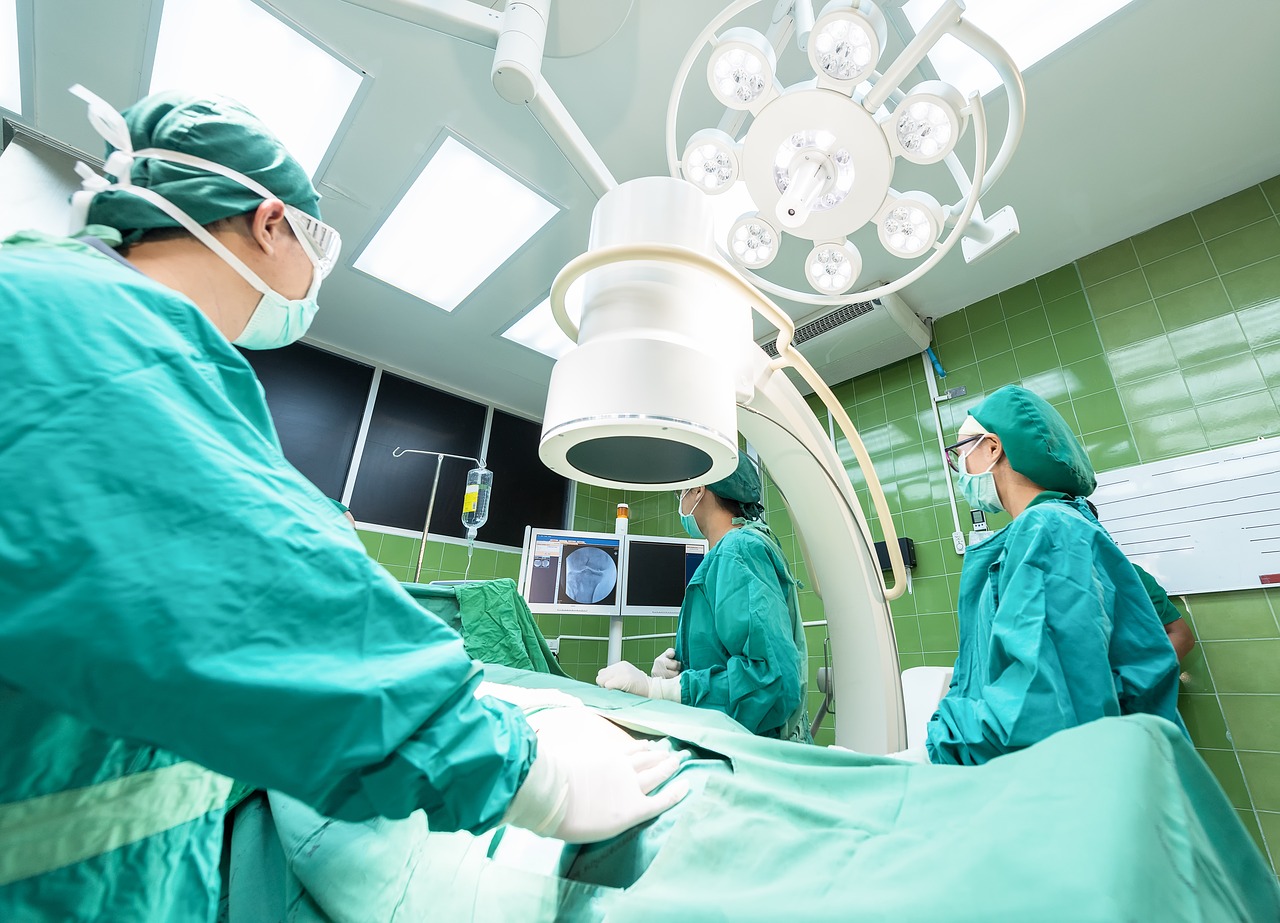
[dropcap]W[/dropcap]annaCry was the first case of a cyberattack that had a major effect on hospitals. The increasing digitization of hospitals (like all areas of society) offers significant opportunities for reducing the cost of health care while making the care provided more effective. However, with digitization comes cybersecurity challenges and these threats must be taken into account in implementing e-health solutions.

The hospital: a highly digitized environment

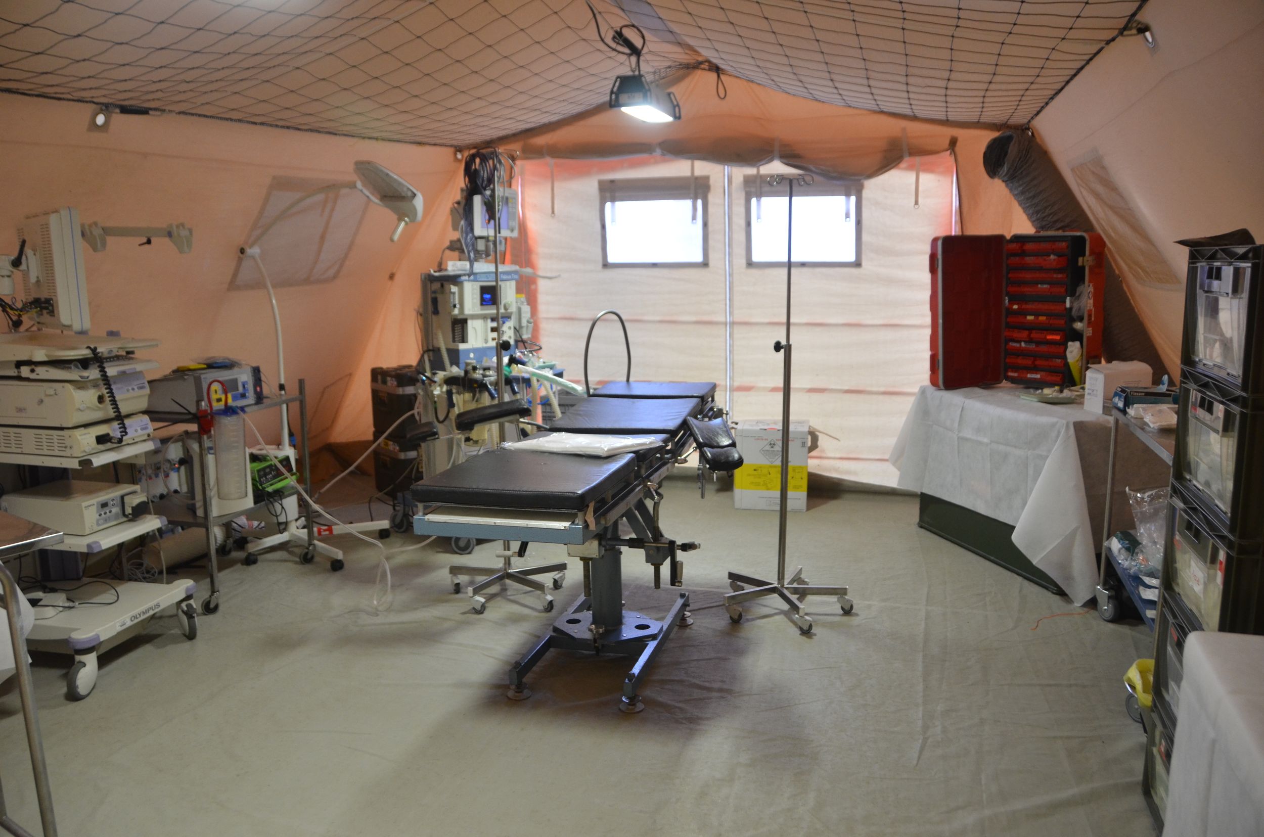
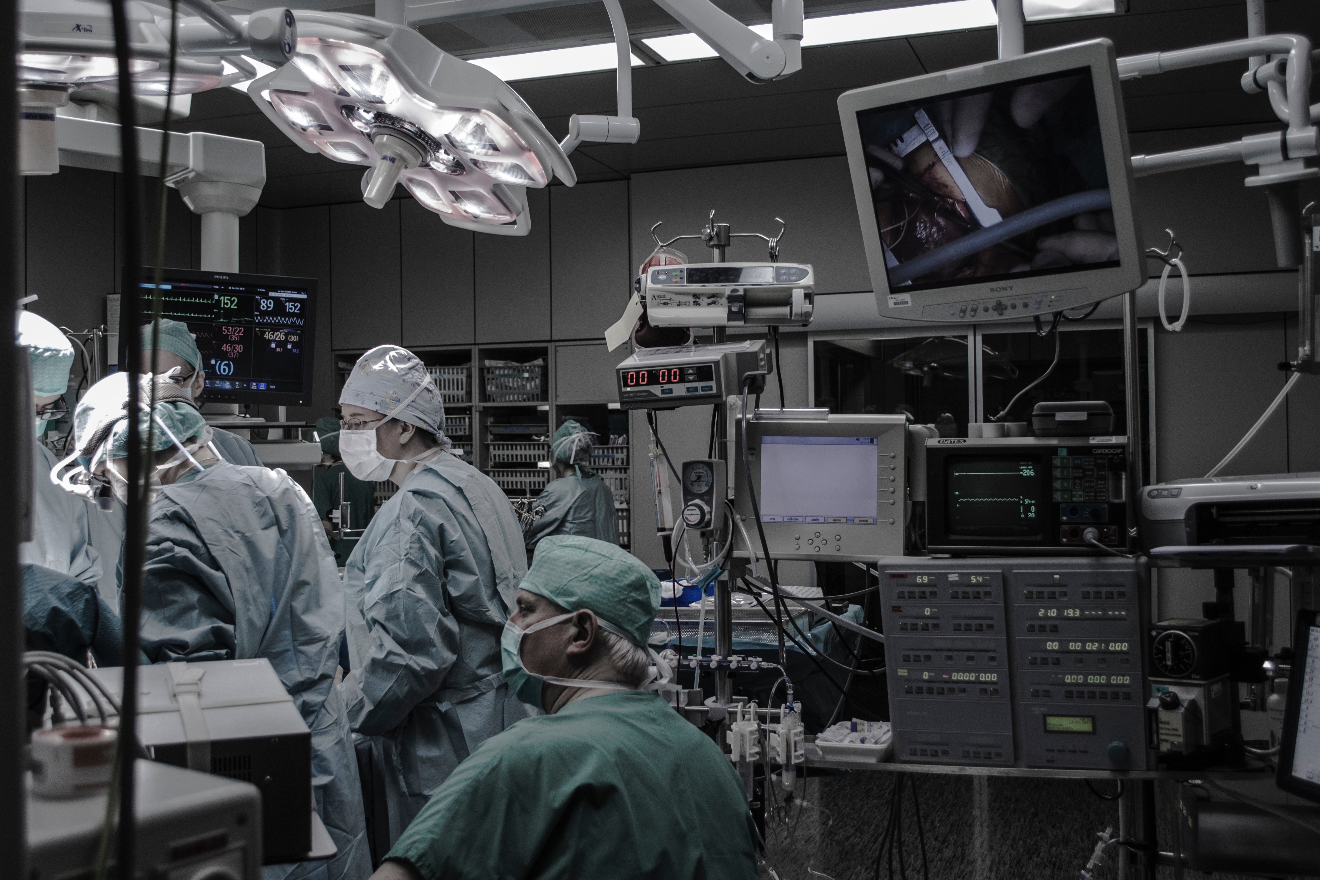
The medical world — and especially hospitals — is a highly digitized environment. This reality first began with management tasks (human resources, room management, planning, etc.) and over the past few years it has grown to include medical equipment (radiology, imaging). Two significant developments have occurred:

  • An increasing number of objects are used in hospitals to collect data or administer medication. This is what is referred to as the Internet of Medical Things (IoMT). The nature of these often-inexpensive objects represents a break with the professional management of conventional medical platforms.
  • More and more of these objects are used outside the hospital, by individuals who are not properly trained to use them. Some of these uncontrolled devices, such as our smartphones, can enter the hospital and interact with medical processes.

From a technical perspective, we are undeniably becoming increasingly dependent on a high-quality digital infrastructure to provide us with quality medical care. This directly affects not just the care provided but also all the related processes (planning, insurance, reimbursement of fees, logistics, etc.). It is particularly difficult to ensure security in these areas, since the conventional development and management technology in information systems is also vulnerable to these attacks. Furthermore, technological advances are based on the increased ability to share, analyze and disseminate information. The number of vulnerabilities is therefore likely to remain high.

From an economic perspective, the rise in healthcare costs is unavoidable. Increased operational efficiency, made possible by computerization, is one of the measures used to prevent costs from rising too high. It is therefore imperative to keep the impacts of cyberattacks in hospital environments to a minimum.

From a legal perspective, the implementation of European personal data protection regulations (GDPR) and the cybersecurity for operators of critical infrastructures (NIS) are imposing new obligations for everyone.

Hospitals are the perfect example of the use of extremely sensitive data demanding confidentiality, integrity (accuracy) and availability (access) to provide care and ensure medical records are properly managed. A medical record is a summary of sensitive, correlated information with separate subsets with varying levels of interest.

A poorly protected environment

Over the past few years there have been cyberattacks that have affected hospital operations. We should note that in many cases, hospitals are just one of the targets of these attacks, since many other organizations are also impacted.

Wannacry is a computer worm that exploits a breakdown in Windows protocol that allows printers and files to be shared. This protocol is used by medical imaging equipment to transfer an image file from a scanner to computers and is used by doctors who meet with patients to make a diagnosis. When imaging equipment is infected by Wannacry through this network protocol, it becomes inoperable, preventing operations and hence endangering patients’ lives.

More generally, much of the medical equipment relies on aging operating systems and old protocol. It is therefore crucial that manufacturers of this equipment become aware of this issue.

The effectiveness of a medical procedure increasingly relies on the ability to connect various tools used by medical staff for the purpose of transferring data (images, prescriptions, etc.) and interacting. Therefore, it is not possible to consider isolating these pieces of equipment. More rigorous access controls must therefore be implemented (which is generally a challenge for organizations, as demonstrated in the study by Deloitte called “Future of Cyber”).

An attack on pacemakers

In addition to the Wannacry incident, it is also necessary to reflect on the communications between medical objects and information systems. Several examples have recently demonstrated the vulnerability of medical objects.

Implants, such as insulin pumps and pacemakers, are vulnerable to computer attacks. Communications between these objects are neither encrypted nor authenticated, meaning that they could be listened to for the purpose of extracting sensitive data. This also means they can receive commands allowing them to be controlled, creating all types of imaginable consequences through changes in their operations.

Other routine medical equipment, like infusion pumps, are also vulnerable to attacks.

New attacks in sight

So far, the attacks that have been revealed have had two main consequences. The first is a denial of service, or the inability to use medical equipment when it is needed and all the potential consequences this entails. Since it is difficult to prevent denial of service attacks, measures must be taken to limit their effects.

The second result is the leak of potentially sensitive information. This leak of information involves the risk of data being added to other databases, for example as data sources for the validation of creditworthiness, used by banks in their decisions to grant or refuse bank loans. This would represent a major setback in protecting our personal data.

We do not have any clear examples of data being falsified, which could be the next step taken by attackers. Data falsification could lead to erroneous prescriptions and therefore to drug diversion. This diversion would allow the author of the crime to receive an immediate profit, which fits with current trends.

What are the solutions?

The first solutions that come to mind are technological ones. Such new solutions do indeed exist which could improve computer security in medical environments.

  • blockchain. This technology can significantly improve data protection by separating the data according to purpose (medical, clerical, insurance, etc.) and by protecting each piece of data individually. It can also log access to manage emergency situations. Current technology is too energy-intensive and must be changed to become more acceptable.
  • Virtualization and cloudification. Outsourcing computer services professionalizes the management of an organization’s digital activities. The scarcity of human resources trained in cybersecurity makes it necessary to rely on external means. The development of cloud services, particularly the concept of a sovereign cloud, must be done in a way that complies with current regulations, particularly the famous GDPR.
  • By Design. Manufacturers of medical objects, software and platforms must take cybersecurity into account during the design phase for their equipment as well as integrating it into the life cycle. This is a major revolution that cannot be carried out in a day. It is therefore necessary to continue protecting older equipment whose initial cost justifies its continued use for decades to come. This is also a revolution for the IT world, which now counts the life span of its software and services in terms of months. While awareness in the area is growing in the industrial world, it must also increase in the medical world.

All these new forms of technology, and others not mentioned here, will never be effective unless the human factor is first taken into account in the hospital, among caregivers, but also patients and visitors. This remains the key to a successful digital transformation of the hospital.

Medical objects must be adapted to their users, generally patients. Besides gadgets like connected watches, better solutions must be found for all objects to make them simpler and easier to use. Confidence in these objects is fundamental and cybersecurity incidents that could restrict their use must be avoided at all costs.

Finally, the role of medical professionals is absolutely fundamental. They must accept the presence of computer technology and recognize that it can make their work easier on a daily basis rather than representing a hindrance. Medical staff must take an interest in cybersecurity issues, receive training in this area and urge suppliers to develop tools adapted to their needs.

[divider style=”normal” top=”20″ bottom=”20″]

Hervé Debar, Head of the Telecoms Networks and Services Department at Télécom SudParis – Institut Mines-Télécom, Université Paris-Saclay

The original version of this article (in French) has been published on The Conversation.

See all articles by Hervé Debar on I’MTech

care pathway

When AI helps predict a patient’s care pathway

Researchers at Mines Saint Etienne are using process mining tools to attempt to describe typical care pathways for patients with a given disease. These models can be used to help doctors predict the next steps for treatment or how a disease will progress.


Will doctors soon be able to anticipate patient complications arising from a disease? Will they be able to determine an entire care pathway in advance for patients with a specific disease? These are the goals of Vincent Augusto and his team at Mines Saint-Étienne. “Based on a patient’s treatment records, their condition at a given moment, and care pathways of similar patients, we’re trying to predict what the next steps will be for the patients,” says Hugo De Oliveira, a PhD student in Health Systems Engineering whose CIFRE thesis is funded by HEVA, a company based in Lyon.

Anticipating how a disease will progress and treatment steps helps limit risks to which the patient is exposed. For people with diabetes — the example used by the researchers in their work — the process is based on detecting weak signals that are precursors of complications as early as possible. For a given patient, the analysis would focus on several years of treatment records and a comparison with other diabetic patients. This would make it possible to determine the patient’s risk of developing renal failure or requiring an amputation related to diabetes.

In order to predict these progressions, the researchers do not rely on personal medical data, such as X-rays or biological analyses. They use medico-administrative data from the national health data system (SNDS). “In 2006, activity-based pricing was put into place,” notes Hugo De Oliveira. With this shift in the principle of funding for healthcare institutions, a large database was created to provide hospitals with the necessary information for reimbursement of treatment. “It’s a very useful database for us, because each line collects information about a patient’s stay: age, sex, care received, primary diagnosis, associated pathologies from which they suffer etc,” says the young researcher.

An entire pathway in one graph

Vincent Augusto’s team is developing algorithms that analyze these large volumes of data. Patients are sorted and put into groups with similar criteria. Different care pathway categories can then be established, each of which groups together several thousands of similar pathways (similar patients, identical complications etc.). In one category — diabetic patients who have undergone amputation for example — the algorithm analyzes all of the steps for the entire group of patients in order to deduce which ones are most characteristic. A graph is produced to represent the typical pathway for this category of patient. It may then be used as a reference to find out whether a patient in the early stages of the disease is following similar steps, and to determine the probability that he/she belongs to this category.

This graph represents the care pathway for patients monitored over an 8-year period who have had a cardiac defibrillator implanted. The part before the implantation can be used to establish statistics for the steps preceding the procedure. The part after the implantation provides information about the future of patients following the implantation.


In this way, the researchers are working on developing longitudinal graphs: each treatment step represents a point on the graph, and the whole graph can be read chronologically: “Doctors can read the graph very easily and determine where the patient is situated in the sequence of steps that characterize his/her pathway,” explains Hugo De Oliveira. The difficulty with this type of data representation comes from its comprehensiveness: “We have to find a way to fit an entire patient pathway into a single line,” says the PhD student. In order to do so, the team chose to use process mining, a data mining and knowledge extraction tool. Machine learning is another such tool.

Process mining helps make care pathway descriptions more effective and easier to read, but it also provides another benefit: it is not a ‘black box’. This characteristic is often encountered in neural network type algorithms. Such algorithms are effective at processing data, but it is impossible to understand the processes that led to the results of the algorithm. Unlike these algorithms, the process mining algorithms used to predict treatment pathways are transparent. “When a patient is characterized by a type of graph, we’re able to understand why by looking at past treatment steps, and studying each graph for the patient’s categories to understand how the algorithm evaluated the pathway,” says Hugo De Oliveira.

Making artificial intelligence applications more transparent is one of the issues brought forth by the working group that produced a French report on AI led by Cédric Villani. The project is also in keeping with the objectives set by the mathematician and member of the French parliament to facilitate AI experimentation for applications, for healthcare in particular. “Our research directly benefits from policies for opening access to health data,” says the PhD student. This access will continue to open up for the researchers, since later on this year they will be able to use the database of the national health insurance cross-scheme system (SNIIRAM): the 1.2 billion healthcare forms contained in the system will be used to improve the algorithms and better identify patient treatment pathways.


fine particles

Fine particles: how can their impact on health be better assessed?

In order to assess the danger posed by fine particles in ambient air, it is crucial to do more than simply take regulatory measurements of their mass in the air. The diversity of their chemical composition means that different toxicological impacts are possible for an equal mass. Chemists at IMT Lille Douai are working on understanding the physicochemical properties of the fine particle components responsible for their adverse biological effects on health. They are developing a new method to indicate health effects, based on measuring the oxidizing potential of these pollutants in order to better identify those which pose risks to our health.


The smaller they are, the greater their danger. That is the rule of thumb to sum up the toxicity of the various types of particles present in the atmosphere. This is based on the ease with which the smallest particles penetrate deep into our lungs and get trapped there. While the size of particles clearly plays a major role in how dangerous they are, the impact of their chemical composition must not be understated. For an equal mass of fine particles in the air, those we breath in Paris are not the same as the ones we breathe in Dunkirk or Grenoble, due to the different nature of the sources which produce them.  And even within the same city the particles we inhale vary greatly depending on where we are located in relation to a road or a factory.

Fine particles are very diverse: they contain hundreds, or even thousands of chemical compounds,” say Laurent Alleman and Esperanza Perdrix, researchers in atmospheric pollution in the department of atmospheric sciences and environmental engineering at IMT Lille Douai. Carboxylic acid, polycyclic aromatic hydrocarbons are just some of the many examples of molecules found in particles in higher or lower proportions. A great number of metals and metalloids can be added to this organic cocktail: copper, iron, arsenic etc., as well as carbon black. The final composition of a fine particle therefore depends on its proximity to sources of each of these ingredients. Copper and antimony, for example, are commonly found in particles near roads, produced by cars when braking, while nickel and lanthanum are typical of fine particles produced from petrochemistry.

Read more on I’MTech: What are fine particles?

Today, only the mass concentration as a function of certain sizes of particles in the air is considered in establishing thresholds for warning the population. For Laurent Alleman and Esperanza Perdrix, it is important to go beyond mass and size to better understand and prevent the health impacts of particles based on their chemical properties.  Each molecule, each chemical species present in a particle has a different toxicity. “When they penetrate our lungs, fine particles break down and release these components,” explains Laurent Alleman. “Depending on their physicochemical properties, these exogenous agents will have a more or less serious aggressive effect on the cells that make up our respiratory system.”

Measuring particles’ oxidizing potential

This aggression mainly takes the form of oxidation chemical reactions in cells: this is oxidative stress. This effect induces deterioration of biological tissue and inflammation, which can lead to different pathological conditions, whether in the respiratory system — asthma, chronic obstructive pulmonary diseases — or throughout the body. Since the chemical components and molecules produced by these stressed cells enter the bloodstream, they also create oxidative stress elsewhere in the body. “That’s why fine particles are also responsible for cardiovascular diseases such as cardiac rhythm disorders,” says Esperanza Perdrix. When it becomes too severe and chronic, oxidative stress can have mutagenic effects by altering DNA and can promote cancer.

For researchers, the scientific challenge is therefore to better assess a fine particle’s ability to cause oxidative stress. At IMT Lille Douai, the approach is to measure this ability in test tubes by determining the resulting production of oxidizing molecules for a specific type of particle. “We don’t directly measure the oxidative stress produced at the cellular level, but rather the fine particle’s potential to cause this stress,” explains Laurent Alleman. As such, the method is less expensive and quicker than a study in a biological environment. Most importantly, “Unlike tests on biological cells, measuring particles’ oxidizing potential is quick and can be automated, while giving us a good enough indication of the oxidative stress that would be produced in the body,” says Esperanza Perdrix. A winning combination, which would make it possible to make oxidizing potential a reference base for the analysis and ongoing, large-scale prevention of the toxicity of fine particles.

To measure the toxicity of fine particles, researchers are finding alternatives to biological analysis.


This approach has already allowed the IMT Lille Douai team to measure the harmfulness of metals. They have found that copper and iron are the chemical elements with the highest oxidizing potential. “Iron reacts with the hydrogen peroxide in the body to produce what we call free radicals: highly reactive chemical species with short lifespans, but very strong oxidizing potential,” explains Laurent Alleman. If the iron provided by the fine particles is not counterbalanced by an antioxidant — such as vitamin C — the radicals formed can break molecular bonds and damage cells.

Researchers caution, however, that, “Measuring oxidizing potential is not a unified method; it’s still in the developmental stages.” It is based on the principle of bringing together the component whose oxidizing potential is to be assessed with an antioxidant, and then measuring the quantity or rate of antioxidant consumed. In order for oxidizing potential to become a reference method, it still has to be become more popular among the scientific community, demonstrate its ability to accurately assess biological oxidative stress produced in vivo, and be standardized.

So for now, the mass concentration of fine particles remains the preferred method. Nevertheless, a growing number of studies are being carried out with the aim of taking account of chemical composition and health aspects. This is reflected in the many disciplines involved in this research. “Toxicological issues bring together a wide variety of fields such as chemistry, physics, biology, medicine, bioinformatics and risk analysis, to name just a few,” says Esperanza Perdrix, who also cites communities other than those with scientific expertise. “This topic extends beyond our disciplinary fields and must also involve environmental groups, citizens, elected officials and others,” she adds. 

Research is ongoing at the international level as well, in particular through MISTRALS, a large-scale meta-program led by CNRS, launched in 2010 for a ten-year period. One of its programs, called ChArMEx, aims to study pollution phenomena in the Mediterranean basin. “Through this program, we’re developing international collaboration to improve methods for measuring oxidizing potential,” explains Laurent Alleman. “We plan to develop an automated tool for measuring oxidizing potential over the next few years, by working together with a number of other countries, especially those in the Mediterranean region such as Crete, Lebanon, Egypt, Turkey etc.”


Also read on I’MTech:

[divider style=”normal” top=”20″ bottom=”20″]

A MOOC to learn all about air pollution

On October 8, IMT launched a MOOC dedicated to air quality, drawing on the expertise of IMT Lille Douai. It presents the main air pollutants and their origin, whether man-made or natural. The MOOC will also provide an overview of the health-related, environmental and economic impacts of air pollution.

[divider style=”normal” top=”20″ bottom=”20″]

healthcare

AI in healthcare for the benefit of individuals and society?

Article written by Christian Roux (Director of Research and Innovation at IMT), Patrick Duvaut (Director of Innovation at IMT), and Eric Vibert (professor at Université Paris-Sud/Université Paris Saclay, and surgeon at Hôpital Paul Brousse (AP-HP) in Villejuif).

[divider style=”normal” top=”20″ bottom=”20″]

How can artificial intelligence be built in such a way that it is humanistic, explainable and ethical? This question is central to discussions about the future of AI technology in healthcare. This technology must provide humans with additional skills without replacing healthcare professionals. Focusing on the concept of “sustainable digital technology,” this article presents the key ideas explained by the authors of a long format report published in French in the Télécom Alumni journal. 


Artificial intelligence (AI) is still in its early stages. Performing single tasks and requiring huge numbers of examples, it lacks a conscience, empathy and common sense. In the medical field, the relationship between patient and augmented caregiver, or virtual medical assistant, is a key issue. In AI, what is needed is a personalized, social learning behavior between machines and patients. There is also a technological and scientific limitation: the lack of explainability in AI. Methods such as deep learning act like black boxes. A single verdict resulting from a process that can be described as “input big data, output results” is not sufficient for practitioners or patients. For the former it is an issue of taking responsibility for supporting, understanding and controlling the patient treatment pathway, while for the latter, it is one of adhering to a treatment and therapy approach, crucial aspects of patients’ involvement in their own care.

However, the greatest challenges facing AI are not technological, but are connected to its use, governance, ethics, etc. Going from “cognitive,” and “social” technologies to decision-making technologies is a major step. It means completely handing over responsibility to digital technology. The methodological bases required for such a transition exist, but putting them to use would have a significant impact on the nature of the coevolution of humans and AI. As far as doctors are concerned, the January 2018 report by the French Medical Council (CNOM) is quite clear: practitioners wish to keep control over decision-making. “Machines must serve man, not control him,” states the report. Machines must be restricted to augmenting decision-making or diagnoses, like IBM Watson. As philosopher and essayist Miguel Benasayag astutely points out, “AI does not ask questions, Man has to ask them.”

Leaving humans in charge of decision-making augmented by AI has become an even more crucial issue in recent years. Since 2016, society has been facing its most serious crisis of confidence in social media and digital service platforms since the dawn of the digital age, with the creation of the World Wide Web in 1990. After nearly 30 years of existence, digital society is going through a paradigm shift. Facing pressure from citizens driven by a crisis of confidence, it is arming itself with new tools, such as the European Data Protection Regulation (GDPR). The age of alienating digital technology, which was free to act as a cognitive, social and political colonizer for three decades, is being replaced by “sustainable digital technology” that puts citizens at the center of the cyber-sphere by giving them control over their data and their digital lives. In short, it is a matter of citizen empowerment.

In healthcare this trend translates into “patient empowerment.” The report by the French Medical Council and the health report for the General Council for the Economy both advocate a new healthcare model in the form of a “health democracy” based on the “6P health”: Preventative, Predictive, Personalized, Participatory, Precision-based, Patient-focused.

“Humanistic AI” for the new treatment pathway

The following extract from the January 2018 CNOM report asserts that healthcare improvements provided by AI must focus on the patient, ethics and building a trust-based relationship between patient and caregiver: “Keeping secrets is the very basis of people’s trust in doctors. This ethical requirement must be incorporated into big data processing when building algorithms.” This is also expressed in the recommendations set forth in Cédric Villani’s report on Artificial Intelligence. Specifically, humanistic artificial intelligence for healthcare and society would include three components to ensure “responsible improvements”: responsibility, measurability and native ethics.

The complexity of medical devices and processes, the large number of parties involved, and the need to instantly access huge quantities of highly secure data require the use of AI as a Trusted Service platforms (AIaaTS), which natively integrate all the virtues of “sustainable digital technology.” AIaaTS is based on a data vault that incorporates the three key aspects of digital cognitive technologies, perception, reasoning and action, and is not limited to only deep or machine learning.

Guarantees of trust, ethics, measurability and responsibility would rely on characteristics that use tools of the new digital society. Native compliance with GDPR, coupled with strong user authentication systems would make it possible to ensure patient safety. The blockchain, with its smart contracts, can also play a role by allowing for enhanced notarization of administrative management and medical procedures. Indicators for ethics and explainability already exist to avoid the black-box effect as much as possible. Other indicators, similar to the Value Based Healthcare model developed by Harvard University Professor Michael Porter, measure the results of AI in healthcare and its added value for the patient.

All these open-ended questions allow us to reflect on the future of AI. The promised cognitive and functional improvements must be truly responsible improvements and must not be made at the expense of alienating individuals and society. If AI can be described as a machine for improvements, humanistic AI is a machine for responsible improvements.


Empathic

AI to assist the elderly

Projets européens H2020Caring and expressive artificial intelligence? This concept that seems to come straight from a man-machine romance like the movie “Her”, is in fact at the heart of a Horizon 2020 project called EMPATHIC. The project aims to develop software for a virtual and customizable coach for assisting the elderly. To learn more, we interviewed the project’s Scientific Director for Télécom SudParis and expert in voice recognition, Dijana Petrovska-Delacretaz.

[divider style=”normal” top=”20″ bottom=”20″]

The original version of this article (in French) was published on the Télécom SudParis website.

[divider style=”normal” top=”20″ bottom=”20″]

What is the goal of the Empathic project?

Dijana Petrovska-Delacretaz: The project’s complete title is “Empathic Expressive Advanced Virtual Coach to Improve Independent Healthy Life-Years of the Elderly”. The goal is to develop a virtually advanced, empathetic and expressive coach to assist the elderly in daily life. This interface equipped with artificial intelligence and featuring several options would be adaptable, customizable and available on several types of media: PC, smartphone or tablet. We want to use a wide range of audiovisual information and interpret it to take the users’ situation into account and try to respond and interact with them in the most suitable way possible to offer them assistance. This requires us to combine several types of technology, such as signal processing and biometrics.

How will you design this AI?

DPD: We will combine signal processing and speech, face and shape recognition using “deep networks” (networks of deep artificial neurons). In other words, we will reproduce our brain’s structure using computer and processor calculations. This new paradigm has become possible thanks to the massive increase in storage and calculation capacities. They allow us to do much more in much less time.

We also use 3D modeling technology to develop avatars with more expressive faces–whether it be a human, cat, robot, hamster or even a dragon–that can be adapted to the user’s preferences to facilitate the dialogue between the user and virtual coach.   The most interesting solution from a biometric and artificial intelligence standpoint is to include as many options as possible: from using the voice and facial expressions to recognizing emotions.  All of these aspects will help the virtual coach recognize its user and have an appropriate dialogue with him or her.

Why not create a robot assistant?

The robot NAO is used at Télécom SudParis in work on recognition and gesture interpretation.

The robot NAO is used at Télécom SudParis in work on recognition and gesture interpretation.

DPD: The software can be fully integrated into a robot like Nao, which is already used here on the HadapTIC and EVIDENT platforms. This is certainly a feasible alternative: rather than have a virtual coach on a computer or smartphone, the coach can be there in person. The advantage with a virtual coach is that it is much easier to bring with me on my tablet when I travel.

In addition, from a security standpoint, a robot like Nao is not allowed access everywhere. We really want to develop a system that is very portable and not bulky. The challenge is to combine all of these types of technology and make them work together so we can interact with the artificial intelligence based on scenarios that are best suited to an elderly individual on different types of devices.

Who is participating in the Empathic project?

DPD: The Universidad del Pais Vasco in Bilbao is coordinating the project and is contributing to the dialogue aspect. We are complementing this aspect by providing the technology for vocal biometrics and emotional and face recognition, as well as the avatar’s physical appearance. The industrial partners involved in the project are Osatek, Tunstall, Intelligent Voice and Acapela. Osatek is the largest Spanish company specializing in connected objects for patient monitoring and home care. Tunstall also specializes in this type of material. Intelligence Voice and Acapela, on the other hand, will primarily provide assistance in voice recognition and speech synthesis. In addition, Osatek and Intelligent Voice will be responsible for ensuring the servers are secure and storing the data. The idea is to create a prototype that they will then be able to use.

Finally, the e-Seniors association, the Seconda Universidad of Italy and the Oslo University Hospital will provide the 300 users-testers to test the EMPATHIC interface throughout the project. This provides us with significant statistical validity.

What are the major steps in the project?

DPD: The project was launched in November 2017 and our research will last three years. We are currently preparing for an initial acquisition stage which take place in the spring of 2018. For this first stage, we will make recordings with a “Wizard of Oz” system in the EVIDENT living lab at Télécom SudParis. With this system, we can simulate the virtual agent with a person who is located in another room in order to collect data on the interaction between the user and the agent. This system allows us to create more complex scenarios that our artificial intelligence will then take into account. This first stage will also provide us with additional audiovisual data and allow us to select the best personifications of virtual coaches to offer to users. I believe this is important.

What are the potential opportunities and applications?

DPD: The research project is intended to produce a prototype. The idea behind having industrial partners is for them to quickly obtain and adapt this prototype to suit their needs. The goal of this project is not to produce a specific innovation, but rather to combine and adapt all these different types of technology so that they reach their potential through specific cases—primarily for assisting the elderly.

Another advantage of EMPATHIC is that it reveals many possible applications in other areas: including video games and social networks.  A lot of people interact with avatars in virtual worlds today–like in the video game Second Life, which is where one of our avatars, Bob the Hawaiian, comes from. EMPATHIC could therefore definitely be adapted outside the medical sector.

Do not confuse virtual and reality

The “Uncanny Valley” is a central concept in artificial intelligence and robotics. It theorizes that if a robot or AI possess a human form (whether physical or virtual), it should not resemble humans too closely–unless this resemblance is perfectly achieved from every angle.  “As soon as there is a strong resemblance with humans, the slightest fault is strange and disturbing. In the end, it becomes a barrier in the interactions with the machine,” explains Patrick Horain, a research engineer at Télécom SudParis, specialized in digital imaging.

Dijana Petrovska-Delacretaz has integrated this aspect and is developing the EMPATHIC coaches accordingly: “It is important to keep this in mind. In general, it is a scientific challenge to make a photo-realistic face that actually looks like a loved one without being able to distinguish the real from the fake. But in the context of our project, it is a problem. It could be disturbing or confusing for an elderly person to interact with an avatar that looks like someone real or even a loved one. It is always preferable to propose virtual coaches that are clearly not human.”

[divider style=”normal” top=”20″ bottom=”20″]


Medcam

Medcam: a high-quality image for laparoscopy

300,000 laparoscopies are performed every year in France. At ten euros per minute spent in the operating room for procedures that last from one to ten hours, any time that can be saved is significant. Medcam helps save precious minutes by reducing the time required to clean the camera used. This makes it possible to schedule one additional patient per operating day. 


It was over a family dinner, which would conclude with making initial sketches, that the idea for Medcam first took shape. That day, Clément, an engineer in fluid mechanics, his sister, an expert in the medical sector, and his brother-in-law Yann, who teaches mechanical engineering, were talking about laparoscopy. This minimally invasive surgical procedure widely performed in digestive, urological and gynecological surgery, is based on inserting a camera in the abdomen. The problem is that condensation, accumulated smoke and blood and visceral fat projections are deposited on the lens and constantly degrade the image. Every ten to fifteen minutes, the surgeon must interrupt the procedure to extract the camera and clean it, which leads to a loss of concentration and wastes time when the camera is removed and reinserted.

One device, three benefits

The solution invented by the brand new SMICES company (Smart Medical Devices) in close collaboration with Dr. Joël Da Broi, a surgeon specialized in visceral and digestive surgery, makes it possible to automatically clean the camera during the procedure. The device, which can be adjusted to fit all camera models, does not in any way interfere with surgeons’ use of the camera or practices in the operating room. But it single-handedly solves three problems: it allows the surgeon to work more comfortably so he/she can remain concentrated on his or her work, it saves time and allows the surgeon to add a patient to the operating schedule.

The start of clinical evaluations after promising tests

Developed in collaboration with the mechatronics platform at IMT Mines Alès, the operational prototype only uses components that already exist in the operation room. Medcam has been successfully tested in real conditions on a cadaver at a university hospital center and will begin preclinical evaluations for CE marking in June 2018 so that it can be marketed in 2019. SMICES will be responsible for manufacturing and distributing Medcam and has set a clear objective: to become the market leader for healthcare institutions in France (300,000 laparoscopies per year) and Italy in its first year, in Europe (over 1 million laparoscopies per year) in four years’ time, and ultimately conquer the world (10 million laparoscopies per year).

BioMica

BioMICA platform: at the cutting edge of medical imaging

Belles histoires, Bouton, CarnotAmong the research platforms at Télécom SudParis, BioMICA has developed bio-imaging applications that have already been approved by the medical field. Airways, its 3D representation software, received funding from Télécom & Société Numérique Carnot Institute.

[divider style=”dotted” top=”20″ bottom=”20″]

The original version of this article (in French) was published on the Télécom SudParis website

[divider style=”dotted” top=”20″ bottom=”20″]


One of the recommendations included in the March 2017 France AI Strategy report was to put artificial intelligence to work to improve medical diagnosis. The BioMICA research platform (which stands for Bio-Medical Imaging & Clinical Applications) has made this goal its mission.

We aim to develop tools that can be used in the clinical setting,” says Catalin Fetita, professor at Télécom SudParis and director of the bio-medical imaging platform. “Our applied research focuses on computer-aided diagnosis involving medical and biological imaging,” he explains. As a specialist in image analysis and processing, Catalin Fetita offers the platform true expertise in the area of medical imaging, particularly in lung imaging.

AirWays, or another way of seeing lungs

AirWays is “image marker” software (like biomarkers in biology). Based on a sequence of lung images taken by a scanner, it extracts as much information as possible for clinicians to assist them in their diagnosis by offering a range of different visualization and classification options. “The quantitative aspect is very important, we do not only want to offer better visual quality,” Catalin Fetita explains. “We offer the possibility of obtaining improved measurements of morphological differences in several areas of the respiratory system at different moments in time. This help clinicians decide which treatment to choose.” In terms of quantified results, the software can detect 95% of stenosis cases, which is the narrowing of bronchial tubes that affects respiratory capacity.

AirWays software uses a graphic grid representation of bronchial tube surfaces after analyzing clinical images and then generates 3D images to view them both “inside and outside” (above, a view of the local bronchial diameter using color coding)This technique allows doctors to plan more effectively for endoscopies and operations that were previously performed by sight.

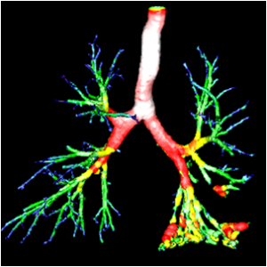
“For now, we have limited ourselves to the diagnosis-analysis aspect, but I would also like to develop a predictive aspect,” says the researcher. This perspective is what motivated Carnot TSN to help finance AirWays in December 2017. “This new budget will help us improve and optimize the software’s interface and increase its computing power to make it a true black box for automatic and synthetic processing,” explains Catalin Fetita, who also hopes to work towards commercializing the software.

A platform for medicine of the future

In addition to its many computer workstations for developing its medical software, the BioMICA platform features two laboratories for biological experimentation. One of the laboratories has a containment level of L1 (any biological agent that is non-pathogenic for humans) and the other is L2 (possible pathogen with low risk). Both will help advance the clinical studies in cellular bio-imaging.

In addition, Catalin Fetita and his team are preparing a virtual reality viewing station to provide a different perspective of the lung tissue analyzed by Airways. “Our platform works thanks to research partnerships and technological transfers,” he explains, “but we can also use it to provide services for clinical studies.”


field hospitals, hôpitaux de campagne

Emergency logistics for field hospitals

European field hospitals, or temporary medical care stations, are standing by and ready to be deployed throughout the world in the event of a major disaster. The HOPICAMP project, of which IMT Mines Alès is one of the partners, works to improve the logistics of these temporary medical centers and develop telemedicine tools and training for health care workers. Their objective is to ensure the emergency medical response is as efficient as possible. The logistical tools developed in the context of this project were successfully tested in Algeria on 14-18 April during the European exercise EU AL SEISMEEX.


European field hospitals, or temporary medical care stations, are standing by and ready to be deployed throughout the world in the event of a major disaster. The HOPICAMP project, of which IMT Mines Alès is one of the partners, works to improve the logistics of these temporary medical centers and develop telemedicine tools and training for health care workers. Their objective is to ensure the emergency medical response is as efficient as possible. The logistical tools developed in the context of this project were successfully tested in Algeria on 14-18 April during the European exercise EU AL SEISMEEX.

Earthquakes, fires, epidemics… Whether the disasters are of natural or human causes, European member states are ready to send resources to Africa, Asia or Oceania to help the affected populations. Field hospitals, temporary and mobile stations where the wounded can receive care, represent a key element in responding to emergencies.

After careful analysis, we realized that the field hospitals could be improved, particularly in terms of research and development,” explains Gilles Dusserre, a researcher at IMT Mines Alès, who works in the area of risk science and emergency logistics. This multidisciplinary field, at the crossroads between information technology, communications, health and computer science, is aimed at improving the understanding of the consequences of natural disasters on humans and the environment. “In the context of the HOPICAMP project, funded by the Single Interministerial Fund (FUI) and conducted in partnership with the University of Nîmes, the SDIS30 and companies CRISE, BEWEIS, H4D and UTILIS, we are working to improve field hospitals, particularly in terms of logistics,” the researcher explains.

Traceability sensors, virtual reality and telemedicine to the rescue of field hospitals

When a field hospital is not being deployed, all the tents and medical equipment are stored in crates, which makes it difficult to ensure the traceability of critical equipment. For example, an electrosurgical unit must never be separated from its specific power cable due to risks of not being able to correctly perform surgical operations in the field. “The logistics operational staff are all working on improving how these items are addressed, identified and updated, whether the hospital is deployed or on standby,” Gilles Dusserre explains. The consortium worked in collaboration with BEWEIS to develop an IT tool for identification and updates as well as for pairing RFID tags, sensors that help ensure the traceability of the equipment.

In addition, once the hospital is deployed, pharmacists, doctors, engineers and logisticians must work in perfect coordination in emergency situations. But how can they be trained in these specific conditions when their workplace has not yet been deployed? “At IMT Mines Alès, we decided to design a serious game and use virtual reality to help train these individuals from very different professions for emergency medicine,” explains Gilles Dusserre. Thanks to virtual reality, the staff can learn to adapt to this unique workplace, in which operating theaters and treatment rooms are right next to living quarters and rest areas in tents spanning several hundred square meters. The serious game, which is being developed, is complementary to the virtual reality technology. It allows each participant to identify the different processes involved in all the professions to ensure optimal coordination during a crisis situation.

Finally, how can the follow-up of patients be ensured when the field hospitals are only present in the affected countries for a limited time period? “During the Ebola epidemic, only a few laboratories in the world were able to identify the disease and offer certain treatments. Telemedicine is therefore essential here,” Gilles Dusserre explains. In addition to proposing specific treatments to certain laboratories, telemedicine also allows a doctor to remotely follow-up with patients, even when the doctor has left the affected area.  “Thanks to the company H4D, we were able to develop a kind of autonomous portable booth that allows us to observe around fifteen laboratory values using sensors and cameras.” These devices remain at the location, providing the local population with access to dermatological, ophthalmological and cardiological examinations through local clinics.

Field-tested solutions

We work with the Fire brigade Association of the Gard region, the French Army and Doctors Without Borders. We believe that all of the work we have done on feedback from the field, logistics, telemedicine and training has been greatly appreciated,” says Gilles Dusserre.

In addition to being accepted by end users, certain tools have been successfully deployed during simulations. “Our traceability solutions for equipment developed in the framework of the HOPICAMP project were tested during the EU AL SEISMEEX Europe-Algeria earthquake deployment exercises in the resuscitation room,” the researcher explains. The exercise, which took place from April 14-18 in the context of a European project funded by DG ECHO, the Directorate-General for European Civil Protection and Humanitarian Aid Operations, simulated the provision of care for victims of a fictional earthquake in Bouira, Algeria. 1,000 individuals were deployed for the 7 participating countries: Algeria, Tunisia, Italy, Portugal, Spain, Poland and France. The field hospitals from the participating countries were brought together to cooperate and ensure the interoperability of the implemented systems, which can only be tested during actual deployment.

La logistique de l’urgence au service des hôpitaux de campagne

The EU-AL SEISMEEX team gathered in front of the facilities for the exercise.


Gilles Dusserre is truly proud to have led a project that contributed to the success of this European simulation exercise. “As a researcher, it is nice to be able to imagine and design a project, see an initial prototype and follow it as it is tested and then deployed in an exercise in a foreign country. I am very proud to see what we designed becoming a reality.”

Diatabase: France’s first diabetes database

The M4P consortium has received the go-ahead from Bpifrance to implement its project to build a clinical diabetes database called Diatabase. The consortium headed by Altran also includes French stakeholders in diabetes care, the companies OpenHealth and Ant’inno, as well as IMT and CEA List. The consortium aims to improve care, study and research for this disease that affects 3.7 million people in France. The M4P project was approved by the Directorate General for Enterprise of the French Ministry for Economy and Finance as part of the Investissements d’Avenir (Investments for the Future) program-National fund for the digital society.


In France, 3.7 million people are being treated for type 1 or type 2 diabetes, which represents 5% of the population. The prevalence of these diseases continues to increase and their complications are a major concern for public health and economic sustainability. Modern health systems produce huge quantities of health data about the disease, both through community practices and hospitals, and additional data is generated outside these systems, by the patients themselves or through connected objects. The potential for using this massive data from multiple sources is far-reaching, especially for advancing knowledge of diabetes, promoting health and well-being of diabetes sufferers and improving care (identifying risk factors, diagnostic support, monitoring the efficacy of treatment etc.)

Supported by a consortium of multidisciplinary experts, and backed by the Directorate General for Enterprise of the French Ministry for Economy and Finance, the M4P project aims to build and make available commercially a multi-source diabetes database, “Diatabase,” comprising data from hospitals and community practices, research centers, connected objects and cross-referenced with the medical-economic databases of the SNDS (National Health Data System).

The project seeks to “improve the lives and care of diabetes sufferers through improved knowledge and sharing of information between various hospital healthcare providers as well as between expert centers and community practices,” says Dr Charpentier, President of CERITD (Center for Study and Research for Improvement of the Treatment of Diabetes) one of the initiators of the project. “With M4P and Diatabase, we aim to promote consistency in providing assistance for an individual within a context of monitoring by interdisciplinary teams and to increase professionals’ knowledge to provide better care,” adds Professor Brigitte Delemer, Head of the Diabetology department at the University Hospital of Reims and Vice-President of the CARéDIAB network which is also involved in the M4P project.

“The analysis of massive volumes of ‘real-life’ data involves overcoming technical hurdles, in particular in terms of interoperability, and will further the understanding of the disease while providing health authorities and manufacturers with tools to monitor the drugs and medical devices used,” says Dr Jean-Yves Robin, Managing Director of OpenHealth, a company specializing in health data analysis which is part of the M4P project.

The M4P is supported by an expert consortium bringing together associations of healthcare professionals active in diabetes, such as CERITD, the CARéDIAB network, the Nutritoring company; private and public organizations specializing in digital technologies with Altran, data analysis with OpenHealth and ANT’inno, associated with CEA List for the semantic analysis and use of unstructured data, and finally, Institut Mines Telecom, which is providing its Teralab platform, a secure accelerator for research projects in AI and data, and its disruptive techniques for preventing data leaks, misuse and falsification based on data watermarking technology as well as methods for processing natural language to reveal new correlations and facilitate prediction and prevention.

“Thanks to the complementary nature of the expertise brought together through this project,  its business-focused approach and consideration of professional practices, M4P is the first example in France of structuring health data and making it available commercially in the interest of the public good,” says Fabrice Mariaud, Director of Programs, Research and Expertise Centers in France for Altran.

The consortium has given itself a period of three years to build Diatabase and to make it available for use, to serve healthcare professionals and patients.

breast cancer

Superpixels for enhanced detection of breast cancer

Deep learning methods are increasingly used to aid medical diagnosis. At IMT Atlantique, Pierre-Henri Conze is taking part in this drive to use artificial intelligence algorithms for healthcare by focusing on breast cancer. His work combines superpixels defined on mammograms and deep neural networks to obtain better detection rates for tumor areas, thereby limiting false positives.


In France, one out of eight women will develop breast cancer in their lives. Every year 50,000 new cases are recorded in the country, a figure which has been on the rise for several years. At the same time, the survival rate has also continued to rise. The five-year survival rate after being diagnosed with breast cancer increased from 80% in 1993 to 87% in 2010. These results can be correlated with a rise in awareness campaigns and screening for breast tumors. Nevertheless, large-scale screening programs still have room for improvement. One of the major limitations of this sort of screening is that it results in far too many false positives, meaning patients must come back for additional testing. This sometimes leads to needless treatment with serious consequences: mastectomy, radiotherapy, chemotherapy etc. “Out of 1,000 participants in a screening, 100 are called back, while on average only 5 are actually affected by breast cancer,” explains Pierre-Henri Conze, a researcher in image processing. The work he carries out at IMT Atlantique in collaboration with Mines ParisTech strives to reduce this number of false positives by using new analysis algorithms for breast X-rays.

The principle is becoming better-known: artificial intelligence tools are used to automatically identify tumors. Computer-aided detection helps radiologists and doctors by identifying masses, one of the main clinical signs of breast cancer. This improves diagnosis and saves time since multiple readings do not then have to be carried out systematically. But it all comes down to the details: how exactly can the software tools be made effective enough to help doctors? Pierre-Henri Conze sums up the issue: “For each pixel of a mammography, we have to be able to tell the doctor if it belongs to a healthy area or a pathological area, and with what degree of certainty.”

But there is a problem: algorithmic processing of each pixel is time-consuming. Pixels are also subject to interference during capture: this is “noise,” like when a picture is taken at night and certain pixels are whited out. This makes it difficult to determine whether an altered pixel is located in a pathological zone or not. The researcher therefore relies on “superpixels.” These are homogenous areas of the image obtained by grouping together neighboring pixels. “By using superpixels, we limit errors related to the noise in the image, while keeping the areas small enough to limit any possible overlapping between healthy and tumor areas,” explains the researcher.

In order to successfully classify the superpixels, the scientists rely on descriptors: information associated with each superpixel to describe it. “The easiest descriptor to imagine is light intensity,” says Pierre-Henri Conze. To generate this information, he uses a certain type of deep neural network, called a “convolutional” neural network. What is their advantage compared to other neural networks? They determine by themselves which descriptors are the most relevant in order to classify superpixels using public mammography databases. Combining superpixels with convolutional neural networks produces especially successful results. “For forms as irregular as tumor masses, this combination strives to identify the boundaries of tumors more effectively than with traditional techniques based on machine learning,” says the researcher.

This research is in line with work by the SePEMeD joint laboratory between IMT Atlantique, the LaTIM laboratory, and the Medecom company, whose focus areas include improving medical data mining. It builds directly on research carried out on recognizing tumors in the liver. “With breast tumors, it was a bit more complicated though, because there are two X-rays per breast, taken at different angles and the body is distorted in each view,” points out Pierre-Henri Conze. One of the challenges was to correlate the two images while accounting for distortions related to the exam. Now the researcher plans to continue his research by adding a new level of complexity: variation over time. His goal is to be able to identify the appearance of masses by comparing different exams performed on the same patient several months apart. The challenge is still the same: to detect malignant tumors as early as possible in order to further improve survival rates for breast cancer patients.

Also read on I’MTech