nose

An “electronic nose” analyzes people’s breath to help sniff out diseases

In partnership with IMT Atlantique, a team of researchers at IMT Lille Douai have developed a device which can measure the level of ammonia in someone’s breath. The aim of the artificial nose is to use this device to create a personalized follow-up care for patients affected by chronic kidney disease.  Eventually, the machine could even allow doctors to detect the disease in undiagnosed people.


This article is part of our dossier “When engineering helps improve healthcare

In the human body, the kidneys’ main role is to remove toxins which are carried in the blood. However, when a person suffers from chronic kidney disease, this filtration function no longer works to the same standard. In France, the disease affects around 5.7 million people and can range from causing a degree of impairment, to terminal. Around 76,000 people are terminally affected in France, which means that their kidneys cannot filter their blood at all.

For these patients, the only options are to either wait for a transplant or face extensive treatment which hugely affects their daily life. It is therefore essential for doctors to be able to detect this silent and progressive disease early enough to slow down the effects. At the moment, doctors use blood or urine tests to identify the disease. But, to make it easier to diagnose, scientists are exploring another route: breath analysis. Studying the substances in the air we breathe out can provide valuable information about a person’s health.

Ammonia: a key element

In order to make this possible, two teams of researchers from IMT Lille Douai and IMT Atlantique have been working in partnership with the nephrology department at the Lille University Hospital. For the past three years, they have been developing a compact and turnkey device for doctors. The device is an ‘electronic nose’, a system made up of several sensors that can measure the specific concentration of a substance in someone’s breath.

In the case of chronic kidney disease, the substance being measured is ammonia. Ammonia is mainly produced by intestinal bacteria and is supposed to be filtered from the body by the kidneys. Previous studies have established a concentration threshold for levels of ammonia in a person’s breath, which doctors can then use to determine the likelihood that a patient has chronic kidney disease.

Ammonia offers some resistance

But how can you measure this compound using a portable device? The scientists used a series of sensors which react in the presence of ammonia, as Caroline Duc, team member and researcher at IMT Lille Douai, explains. “The sensors are made from two electrodes with a sensitive surface placed on top of them. The resistance of this surface varies depending on the amount of ammonia present”. When used, the device’s ability to resist electrical current increases when ammonia is present and returns to its initial state when the ammonia disappears. This therefore allows scientists to measure the level of the molecule in a patient’s breath.

Additionally, each sensor in the artificial nose has a unique composition. As Caroline Duc points out, “it is very complicated to use a material whose resistance varies when one type of gas is present”. This is why scientists decided to increase the accuracy of the analysis by combining several different sensors which have different responses to ammonia.

During the tests, the electronic nose was periodically exposed to a person’s breath. This resulted in an increase in the resistance when ammonia was present, and a decrease when the sensors were no longer exposed to the exhaled air.  Several factors were then measured and analyzed using statistical processing algorithms.

These algorithms rely on tools such as machine learning. The only difference with this case was that here, these tools were applied using small amounts of data and  supervised learning to categorize the different types of breath. In other words, the algorithms were taught using a dataset that had already classified breath which belongs to a healthy, ill or ‘uncertain’ individual. New profiles were then passed through this algorithm, so that they could be classed into these three categories.

Breath: a complex compound

To date, the first prototype developed is around 15cm in length and contains between 10 and 13 sensors.  The device was tested in a laboratory using artificial breath, which allowed the teams to verify that it could distinguish a healthy individual from someone who was ill, using the criteria defined in up-to-date scientific literature.  Then, experiments were carried out in clinics with patients suffering from chronic kidney disease. The idea was to measure the concentration of ammonia in their breath, before and after dialysis. The results demonstrated that there was a reduction in ammonia after the treatment.

However, they also highlighted the limitations of using a single marker. Before dialysis, some patients had levels of ammonia that were similar to those of a healthy person. Measuring the amount of ammonia in a person’s breath did not give a reliable diagnosis for chronic kidney disease.  This has caused scientists to launch a new study in order to identify other biomarkers characteristic of the disease.

More generally, this reflects the difficulty of conducting a reliable breath analysis, which can include both the air we breathe in as well as out. “Initially, I think clinical breath analysis will be developed for personalized patient follow-up care, rather than for diagnosis”, says Caroline Duc. For example, by measuring how a patient’s ammonia levels change in response to medication, doctors will be able to monitor the effectiveness of a treatment and then adapt it based on the results.

What is the future of follow-up care for other illnesses?

Researchers at IMT Lille Douai will continue to work on improving the electronic nose. At the moment, patients’ breath is collected and sealed in an airtight bag and is then analyzed in a laboratory. Consequently, the team’s aim is to develop a functional and completely autonomous prototype which would give doctors real-time results. However, this raises several new issues, such as the study of fluids, controlling the speed that the air is exhaled, etc. As well as this, to improve their data analysis, Caroline Duc and her colleagues have started a partnership with researchers who specialize in data handling at Télécom SudParis.

Moreover, the team is involved in a European-wide project which aims to identify biomarkers for lung cancer, and then create a multi-sensor system that is specifically designed to detect these substances. IMT Lille Douai’s expertise will be especially useful for this second objective.

This electronic nose, capable of sniffing out illnesses such as chronic kidney disease, is therefore still in the early stages of development, and needs a lot of work before it can really be used by doctors and their patients. But doctors are waiting with bated breath; in several years’ time it could be a breathtaking medical innovation!

Article written (in French) for I’MTech by Bastien Conteras

professional

When healthcare professionals form communities through digital technology

Digital technology is shaking up the healthcare world. Among its other uses, it can help break isolation and facilitate online interactions in both the private and professional spheres. Can these virtual interactions help form a collective structure and community for individuals whose occupations involve isolation and distance from their peers? Nicolas Jullien, a researcher in economics at IMT Atlantique, looks at two professional groups, non-hospital doctors and home care workers, to outline the trends of these new digital connections between practices.


On the Twitter social network, doctors interact using a bot with the hashtag #DocTocToc. These interactions include all kinds of professional questions, requests for details about a diagnosis, advice or medical trivia. In existence since 2012, this channel for mutual assistance appears to be effective. With rapid interactions and reactive responses, the messages pour in minutes after a question is asked. None of this comes as a surprise for researcher Nicolas Jullien: “At a time when we hear that digital technology is causing structures to fall apart, to what extent does it also contribute to the emergence and organization of new communities?

Doctors–and healthcare professionals in general–are increasingly connected and have a greater voice. On Twitter, some star doctors have thousands of followers: 28,900 for Baptiste Beaulieu, a family doctor, novelist and formerly a radio commentator on France Inter; 25,200 for medical intern and cartoonist @ViedeCarabin; and nearly 9,900 for Jean-Jacques Fraslin, family doctor and author of an op-ed piece denouncing alternative medicine. Until now, few studies had been conducted on these new online communities of practice. Under what conditions do they emerge? What are the constraints involved in their development? What challenges do they face?

New forms of collective action

For several years, Nicolas Jullien and his colleagues have been studying the structure and development of online communities. These forms of collective action, which exist in the popular imagination as the prisoner’s dilemma, question the way action is taken: the classic dilemma involves two suspects arrested by the police and isolated for interrogation. They are then given three possible outcomes: the one can denounce the other and be released. In this case, the accomplice receives the maximum sentence. They can plead guilty or denounce each other and receive a more lenient sentence, or they can both deny their wrongdoings and receive the minimum sentence. Although it would be to the individuals’ advantage to take collective action–and despite an awareness of this fact–it is not always done. In the absence of dialogue, individuals seek to maximize their individual interests. Is it in fact possible for digital platforms to facilitate the coordination of these individual interests? Can they be used to create collective projects for sharing, knowledge and mutual assistance, particularly in the professional sphere, with projects like Wikipedia and open source software? These professional social networks represent a new field of exploration for researchers.

Read on I’MTech: Digital commons:  individual interests to serve a community

We decided to focus on two professional groups, doctors and home care workers, which are both involved in health and service relationships, but are diametrically opposed in terms of qualifications. The COAGUL project, funded by the Brittany Region, analyzes the relationships established in each of these professional groups through online interactions. We are conducting these studies with Christèle Dondeyne’s team at Université de Bretagne Occidentale”, Nicolas Jullien explains. These interactions can be linked to technical problems and uncertainties (diagnoses, ways of performing a procedure), ethical and legal issues (especially related to terms and conditions of employment contracts and relations with the health insurance system), employment or working conditions (amount of time spent providing care at a home, discussions on home care worker tasks that go beyond basic health procedures). Isolation therefore favors the emergence of communities of practice. “Digital technology offers access to tools, platforms and means of coordinating work and the activities of professionals. These communities develop autonomously and spontaneously,” the researchers add.

So how can a profession be carried out online? The researchers are currently conducting work that is exploratory and necessarily qualitative. For the first phase of their study, they identified 20 stakeholders mobilized on the internet in order to determine the ties of cooperation and solidarity that are being created on social networks, forums and dedicated websites by collecting their interactions.  Where do people go? Why do they stay there? “We have observed that usage practices vary according to profession. While home care workers interact more on Facebook, with several groups of thousands of people and a dozen messages per day, family doctors prefer to interact on Twitter and form more personal networks,” Nicolas Jullien explains. “This qualitative method allows us to understand what lies behind the views people share.  Because in these shifting groups, tensions arise related to position and each individual’s experiences,” the researcher explains. For the second phase of the study, researchers will conduct a series of interviews with local professional groups. The long-term objective is to compare various motivations for action and means of interaction from the two different professions.

On a wider scale, behind these digital issues, researchers are seeking to analyze the capacity of these groups to collectively produce knowledge. “In the past, we worked on the Wikipedia model, free online encyclopedia software that brings together nearly 400,000 contributors per month. This collective action is the most extensive that has ever been accomplished. It has seen massive success–that is unexpected and lasting–in producing knowledge online,” Nicolas Jullien explains.

But although contributors participate on a volunteer basis, the rules for contributions are becoming increasingly strict, for example through moderation or based on topics that have already been created. “The contribution is what is regulated in these communities, not the knowledge,” the researcher adds. “Verifying the contribution is what takes time and, for communities of practice, responding. An increase in participants and messages brings with it a greater need to limit noise, i.e. irrelevant comments.” With intellectual challenges, access to peers, and the ability to have contributions viewed by others, the digital routine of daily professional acts has the potential to shake up communities of practice and support the development of new forms of professional solidarity.

Article written (in French) by Anne-Sophie Boutaud, for I’MTech.

cerveau, brain

Imaging to help people with brain injuries

People with brain injuries have complex cognitive and neurobiological processes. This is the case for people who have suffered a stroke, or who are in a minimally conscious state and close to a vegetative state. At IMT Mines Alès, Gérard Dray is working on new technology involving neuroimaging and statistical learning. This research means that we can improve how we observe patients’ brain activity. Ultimately, his studies could greatly help to rehabilitate trauma patients. 


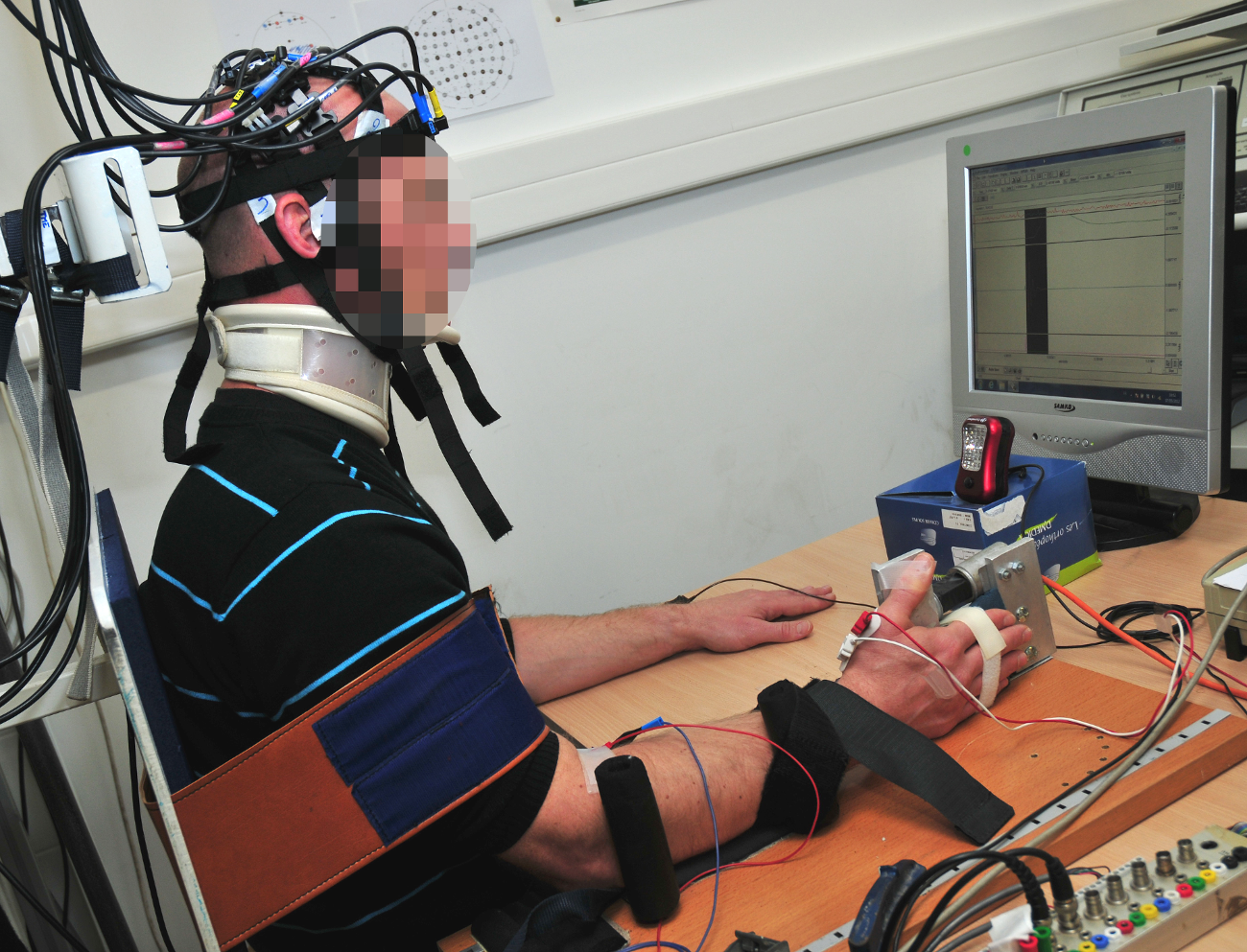
As neuroimaging technology is becoming more effective, the brain is slowly losing its mystery; and as our ability to observe what is happening inside this organ becomes more accurate, we are opening up numerous possibilities, notably in medicine.  For several years, at IMT Mines Alès, Gérard Dray has been working on new tools to detect brain activity.  More precisely, he is aiming to improve how we record and understand the brain signals recorded by techniques such as electroencephalography (EEG) or infrared spectroscopy (NIRS). In partnership with the University of Montpellier’s research center EuroMov, and Montpellier and Nîmes University Hospitals, Dray is putting his research into application in order to support patients who have suffered heavy brain damage.

Read on I’MTech Technology that decrypts the way our brain works

This is notably the case of stroke victims; a part of their brain does not get enough blood supply from the circulatory system and therefore becomes necrotic. The neurons in this part of the brain die and the patient can lose certain motor functions in their legs or arms. However, this disability is not necessarily permanent. Appropriate rehabilitation can mean that stroke victims regain a part of their motor ability. “This is possible thanks to the plasticity of the brain, which allows the brain to move functions stored in the necrotic zone into a healthy part of the brain,” explains Gérard Dray.

Towards Post-Stroke Rehabilitation

In practice, this transfer happens thanks to rehabilitation sessions. Over several months, a stroke victim who has lost their motor skills is asked to imagine moving the part of their body that they are unable to move. In the first few sessions, a therapist guides the movement of the patient. The patient’s brain begins to associate the request for movement to the feeling of the limb moving; and gradually it recreates these neural connections in a healthy area of the brain. “These therapies are recent, less than 20 years old,” points out the researcher at IMT Mines Alès. However, although they have already proven that they work, they still have several limitations that Dray and his team are trying to overcome.

One of the problems with these therapies is the great uncertainty as to the patient’s involvement. When the therapist moves the limb of the victim and asks them to think about moving it, there is no guarantee that they are doing the exercise correctly. If the patient’s thoughts are not synchronized with their movement, then their rehabilitation will be much slower, and may even become ineffective in some cases. By using neuroimaging, researchers want to ensure that the patient is using their brain correctly and is not just being passive during a kinesiotherapy session. But the researchers want to go one step further. By knowing when the patient is thinking about lifting their arm or leg, it is possible to make a part of rehabilitation autonomous.

With our partners, we have developed a device that detects brain signals, and is connected to an automatic glove,” describes Gérard Dray. “When we detect that the patient is thinking about lifting their arm, the glove carries out the associated movement.” The researcher warns that this cannot and should not replace sessions with a therapist, as these are essential for the patient to understand the rehabilitation system.  However, the device allows the victim to complete the exercises in the sessions by themselves, which speeds up the transfer of brain functions towards a healthy zone.  Like after fracture, stroke patients will often have to go through physiotherapy sessions both at the hospital and at home by themselves.

Un gant connecté à un système de détection de l'activité du cerveau peut aider à la rééducation post-AVC.

A glove which is connected to a brain activity detection system can help post-stroke rehabilitation.


The main task for this device is being able to detect the brain signals associated with the movement of the limb.  When observing brain activity, the imaging tools record a constellation of signals associated with all the background activities managed by the brain. The neuronal signal which causes the arm to move gets lost in the crowd of background signals.  In order to isolate it, the researchers use statistical learning tools. The patients are first asked to carry out guided and supervised motor actions, while their neural activity is recorded. Then, they move freely during several sessions, while being monitored by EEG or NIRS technology. Once sufficient data has been collected, the algorithms can categorize the signals by action and can therefore deduce, through real-time neuroimaging, if the patient is in the process of trying to move their arm or not.

In partnership with Montpellier University Hospital, the first clinical trial with the device was carried out on 20 patients. The results were used to test the device. “Although the results are positive, we are still not completely satisfied with them,” admits Dray. “The algorithms only detected the patients’ intention to move their arm in 80% of cases. This means that in two out of ten times, the patient thinks about doing it without us being able to record their thoughts using neuroimaging.” To improve these detection rates, researchers are working on numerous algorithms which categorize brain activity.  “Notably, we are trying to couple imagery techniques with techniques that can detect fainter signals,” he continues.

Detecting Consciousness After Head Trauma

The improvement in these brain activity detection tools is not only useful for post-stroke rehabilitation. The IMT Mines Alès team uses the technology that they have developed on people who have suffered head trauma and whose state of consciousness has been altered. After an accident, a victim who is not responsive, but whose respiratory and circulatory functions are in good condition, can be in several different states. They can be in either a total and normal state of consciousness, a coma, a vegetative state, a state of minimal consciousness, or have locked-in syndrome. “These different states are characterized by two factors, consciousness and being awake,” summarizes Dray. In a normal state, we are both awake and conscious. However, a person who is in a coma is neither awake nor conscious. A person is a vegetative state is awake but not conscious of their surroundings.

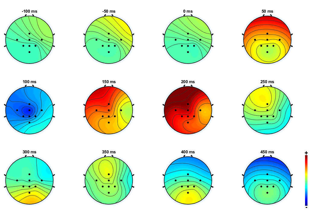
According to their different states, patients receive different types of care and have different prospects of recovery. The huge difficulty for doctors is being able to identify patients who are awake without being responsive, but whose state of consciousness is not yet gone. “With these people there is a hope that their state of consciousness will be able to return to normal,” explains the researcher. However, the patient’s state of consciousness is sometimes very weak, and we have to detect it using high-quality neuroimaging tools. For this, Gérard Dray and his team use EEG paired with sound stimuli. He explains the process. “We speak to the person and explain to them that we are going to play them a series of signals which have deep frequencies, in between these signals there will be high-pitched frequencies. We ask them to count the high-pitched frequencies. Their brain will react to each sound. However, when they are played a high-pitched sound, their cognitive response will be more important, as these are the signals which the brain will remember. More precisely, a wave called P300 is generated when we are innervated. In the case of the high-pitched sounds, the patient’s brain will generate this wave in an important way.”

Temporal monitoring of brain activity after an auditory stimulus using an EEG device.


The patients who still have a state of consciousness will produce a normal EEG in response to the exercise, despite not being able to communicate or move. However, a victim who is in a vegetative state will not respond to the stimuli. The results of these first clinical trials carried out on patients who had experienced head trauma are currently being analyzed. The first bits of feedback are promising for the researchers, who have already managed to detect differences in P300 wave generation. “Our work is only just beginning,” states Gérard Dray. “In 2015, we started our research on the detection of consciousness, and it’s a very recent field.” With increasing progress in neuroimaging techniques and learning tools, this is an entire field of neurology that is about to undergo major advances.


visual impairments

Virtual reality improving the comfort of people with visual impairments

People suffering from glaucoma or retinitis pigmentosa develop increased sensitivity to light and gradually lose their peripheral vision. These two symptoms cause discomfort in everyday life and limit the social activity of the people affected. The AUREVI research project involving IMT Mines Alès aims to improve the quality of life of visually-impaired people with the help of a virtual reality headset.


Retinitis pigmentosa and glaucoma are degenerative diseases of the eye. While they have different causes, they result in similar symptoms: increased sensitivity to changes in light and gradual loss of peripheral vision. The AUREVI research project was launched in 2013 in order to help overcome these deficiencies. Over 6 years, the project has brought together researchers from IMT Mines Alès and Institut ARAMAV in Nîmes, which specializes in rehabilitation and physiotherapy for people with visual impairments. Together, the two centers are developing a virtual reality-based solution to improve the daily lives of patients with retinitis pigmentosa or glaucoma.

“For these patients, any light source can cause discomfort” explains Isabelle Marc, a researcher in image processing at IMT Mines Alès, working on the AUREVI project. A computer screen, for example, can dazzle them. When walking around outdoors, the changes in light between shady and bright areas, or even breaks in the clouds, can be violently dazzling. “For visually impaired people, it takes much longer for the eye to adjust to different levels of light than it does for healthy people” the researcher adds. “While it usually takes a few seconds before we can open our eyes after being dazzled or to be able to see better in a shady area, these patients need several tens of seconds, sometimes several minutes.

Controlling light

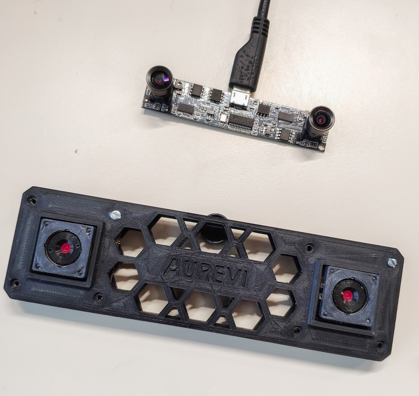
With the help of a virtual reality headset, the AUREVI team offers greater control over light levels for visually impaired people with retinitis or glaucoma. Cameras display the image which would normally be seen by the eyes on the screens of the headset. When there is a sudden change in the light, image processing algorithms alter the brightness of the image in order to keep it constant in the patient’s eyes. For the researchers, the main difficulty with this tool is the delay. “We would like it to be effective in real time. We are aiming for the shortest delay between what appears on the screen of the headset and what the user really sees” says Isabelle Marc. The team is therefore using logarithmic cameras, which record HDR (High Dynamic Range) images directly, thus reducing the processing time.

The headset is designed to replace the dark glasses usually worn by people with this type of pathology. “It’s a pair of adaptive dark sunglasses. The shade varies pixel by pixel” Isabelle Marc explains. An advantage of this tool is that it can be calibrated to suit each patient. Depending on the stage of the retinitis or glaucoma, the level of sensitivity will be different. This can be accounted for in the way the images are processed. To do so, scientists have developed a specific test for evaluating the degree of light a person can bear. “This test could be used to configure the tool and adapt it optimally for each user” says the researcher.

The first clinical trials of the headset began on fifteen people in 2016. The initial goal was to measure the light levels considered as comfortable by each person, and to gather feedback on the comfort of the tool, before looking at evaluating the service provided to people with visual impairments. For this, the researchers create variations in the brightness of a screen for patients wearing a headset, who then give their feedback. Isabelle Marc reports that “the initial feedback from patients shows that they prefer the headset over other tools for controlling light levels”. However, the testers also commented on the bulk of the tool. “For now, we are working with the large headsets available on the market, which are not designed to be worn when you are walking around” the researcher concedes. “We are currently looking for industrial partners who could help us make the shift to a pre-series prototype more suitable for walking around with.

Showing what patients can’t see

Being able to control light levels is a major improvement in terms of visual comfort for patients, but the researchers want to take things even further. The AUREVI project aims to compensate for another symptom caused by glaucoma and retinitis: loss of stereo vision. Patients gradually lose degrees in their visual field, reaching 2 or 3 degrees at around 60 years old, or even full blindness. Before this last stage comes an important step in the progression of the handicap, as Isabelle Marc describes: “Once the vision goes below 20 degrees of the visual field, the images in the eye no longer cross over, and the brain cannot reconstruct the 3D information.

Par des techniques de traitement d'image, le projet AUREVI veut donner aux personnes malvoyantes des indications sur les obstacles à proximité.

Using image processing techniques, the AUREVI project hopes to give people with visual impairments indications about nearby objects


Without stereoscopic vision, the patient can no longer perceive depth of field. One of the future steps of the project will be to incorporate a feature into the headset to compensate for this deficiency in three-dimensional vision. The researchers are working on methods for communicating information on depth. They are currently looking at the idea of displaying color codes. A close object would be colored red, for example, and a far object blue. As well as improving comfort, this feature would also provide greater safety. Patients suffering from an advanced stage of glaucoma or retinitis do not see objects above their head which could hurt them, nor do they see those at their feet which are a tripping hazard.

Losing information about their surroundings gives people with visual impairments the feeling of being in danger, which increases as the symptoms get worse. Combined with an increasing discomfort with changes in light levels, this fear can often lead to social exclusion. “Patients tend to go out less, especially outdoors” notes Isabelle Marc. “On a professional level, their refusal to participate in activities outside of work with their colleagues is often misunderstood. They may still have good sight for reading, for example, and so people with normal sight may have a hard time understanding their handicap. Therefore, their social circle gradually shrinks. The headset developed by the AUREVI project is an opportunity to improve social integration for people with visual impairments. For this reason, it receives financial support from several companies as part of their disabilities and diversity missions, in particular: Sopra Steria, Orano, Crédit Agricole and Thalès. The researchers rely on this aid for people with disabilities in developing their project.

maladie chronique, chronic disease

Chronic disease: what does the Internet really change in patients’ lives?

For the first time, a study has assessed the impact of digital technology on the lives of patients with chronic diseases. It was conducted by the ICA patient association collective, in partnership with researchers from the Smart Objects and Social Networks chair at Institut Mines-Télécom Business School. The study provides a portrait of the benefits and limitations perceived by chronically ill people for three technologies: the Internet, mobile applications and smart objects. Multiple factors were evaluated, such as the quality of the relationship with the physician, the degree of expertise, the patient’s level of incapacitation and their quality of life.


Internet research has become an automatic reflex to learn about any disease. From the common cold to the rarest diseases, patients find information about their cases through more or less specialized sites. Scientific publications have already shown that social networks and health forums are especially used by patients when they are diagnosed. However, the true usefulness of the Internet, apps or smart objects for patients remains unclear. To gain a better understanding of how new technology helps patients, the Impatients, Chroniques & Associés collective (ICA) contacted the Smart Objects and Social Networks Chair at Institut Mines-Télécom Business School. The study, conducted between February and July 2018, focused on people living with chronic disease and their use of digital technology. The results were presented on February 20, 2019 at the Cité des Sciences et de l’Industrie in Paris.

More than 1,013 patients completed the questionnaire designed by the researchers. The data collected on technology usage shows that, overall, patients are not very attracted by smart objects. 71.8% of respondents reported that they used the Internet only, 1 to 3 times a month. 19.3% said they used both the Internet and mobile applications on a weekly basis. Only 8.9% use smart objects in addition to the Internet and apps.

Read on I’MTech Healthcare: what makes some connected objects a success and others a flop?

The study therefore shows that uses are very different and that a certain proportion of patients are characterized by the “multi-technology” category. However, “contrary to what we might think, the third group comprising the most connected respondents is not necessarily made up of the youngest people,” indicates Christine Balagué, holder of the Smart Objects and Social Networks chair. In the 25-34 age group, the study found “almost no difference between the three technology use groups (20% of each use group is in this age group)“. The desire for digital health solutions is therefore not a generational issue.

Digital technology: a relative benefit for patients?

The specificity of the study is that it cross-references the use of digital technology (Internet, mobile applications and smart objects) with standard variables in publications that characterize patients’ behavior towards their health. This comparison revealed a new result: the patients who use technology the most are on average no more knowledgeable about their disease than patients who are not very connected. They are also no more efficient in their ability to adopt preventive behavior related to their disease.

On the other hand, the more connected patients are, the greater their ability is to take action in the management of their disease,” says Christine Balagué. Patients in the most connected category believe they are better able to make preventive decisions and to reassure themselves about their condition. However, technology has little impact on the patient-doctor relationship. “The benefit is relative: there is a difference between the benefit perceived by the patient and the reality of what digital tools provide,” concludes Christine Balagué.

Some of the criteria measured by the researchers nevertheless show a correlation with the degree of use of technology and the use of several technology devices. This is the case, for example, with patient empowerment. Notably, the most connected patients are also those who most frequently take the initiative to ask their doctor for information or give their opinion about treatment. These patients also report being most involved by the doctor in medical care. On this point, the study concludes that:

“The use of technology[…] does not change the self-perception of chronically ill patients, who all feel equally knowledgeable about their disease regardless of their use of digital technology. On the other hand, access to this information may subtly change their position in their interactions with the medical and nursing teams, leading to a more positive perception of their ability to play a role in decisions concerning their health.”

The flip side of the coin

Although information found on the Internet offers genuine benefits in the relationship with the medical profession, the use of technologies also has some negative effects, according to patient feedback. 45% believe that the use of digital technology has negative emotional consequences. “Patients find that the Internet reminds them of the disease on a daily basis, and that this increases stress and anxiety,” says Christine Balagué. This result may be linked to the type of use among the chronically ill. The vast majority of them generally search for stories from other people with similar pathologies, which frequently exposes them to the experiences of other patients and their relatives.

Personal stories are considered the most reliable source of information by patients, ahead of content provided by health professionals and patient associations, a fact due to the large, and unequal, amount of information available. Three quarters of respondents indicated that it is difficult to identify and choose reliable information. This sense of mistrust is underlined by other data collected by the researchers during the questionnaire: “71% believe that the Internet is likely to induce self-diagnosis errors.” In addition, a certain proportion of patients (48%) also express mistrust of the privacy of certain mobile sites and applications. This point highlights the challenge for applications and websites to improve the transparency of the use of personal data and respect for privacy, in order to gain their trust.

Read on I’MTech Ethical algorithms in health: a technological and societal challenge

The future development of dedicated web services and patient usage is an issue that researchers want to address. “We want to continue this work of collecting experiences to evaluate changes in use over time,” says Christine Balagué. The continuation of this work will also integrate other developing uses, such as telemedicine and its impact on patients’ quality of life. Finally, the researchers are also considering taking an interest in the other side: the doctors’ side. How do practitioners use digital technologies in their practice? What are the benefits in the relationship with the patient? By combining the results from patient and physician studies, the aim will be to obtain the most accurate portrait possible of patient-physician relationships and of treatment processes in the era of hyperconnectivity.


Digital twins in the health sector: mirage or reality?

Digital twins, which are already well established in industry, are becoming increasingly present in the health sector. There is a wide range of potential applications for both diagnosis and treatment, but the technology is mostly still in the research phase.


The health sector is currently undergoing digital transition with a view to developing “4P” treatment: personalized, predictive, preventive and participative. Digital simulation is a key tool in these changes. It consists in creating a model of an object, process or physical process on which different hypotheses can be tested. Today, patients are treated when they fall sick based on clinical studies of tens or, at best, thousands of other people, with no real consideration of the individual’s personal characteristics. Will each person one day have their own digital twin to allow prediction of the development of acute or chronic diseases based on their genetic profile and environmental factors, and anticipation of their response to different treatments?

“There are a lots of hopes and dreams built on this idea,” admits Stéphane Avril, Researcher at Mines Saint-Étienne. “The profession of doctor or surgeon is currently practiced as an art on the strength of experience acquired over time. The idea is that a program could combine the experience of a thousand doctors, but in reality there is a huge amount of work still to do to create equations for and integrate non-mathematical knowledge and skills in very different fields such as immunology and the cardio-vascular system.”  We are still a long way from simulating an entire human being. “But in certain fields, digital twins provide excellent predictions that are even better than those of a practitioner,” adds Stéphane Avril.

From imaging to the operating room

Biomechanics, for example, is a field of research that lends itself very well to digital simulation. Stéphane Avril’s research addresses aortic aneurysms. 3D digital twins of the affected area are developed based on medical images. The approach has led to the creation of a start-up called Predisurge, whose software allows the creation of individual endoprostheses. “The FDA [American Food and Drug Administration] is encouraging the use of digital simulation for validating the market entry of prostheses, both orthopedic and vascular,” explains the researcher from St Etienne.

Digital simulation also helps surgeons prepare for operations because the software provides predictions on the insertion and effects of these endoprostheses once in place, as well as simulating any pre-surgical complications that could arise. “This technique is currently still in the testing and validation phase, but it could have a very promising impact on reducing surgery time and complications,” stresses Stéphane Avril. The team at Mines Saint-Étienne is currently working on improving our understanding of the properties of the aortic wall using four-dimensional MRI and mechanical tests on aneurysm tissue removed during the insertion of prostheses. The idea is to validate a digital twin designed using 4D MRI images, which could predict the future rupture or stability of an aneurysm and indicate the need for surgery or not.

Read more on I’MTech: A digital twin of the aorta to prevent Aneurysm rupture

Catalin Fetita, a researcher at Télécom SudParis, also uses digital simulation in the field of imaging, but this time in the case of air-borne transmission alongside pulmonary parenchyma analysis. The aim of her work is to obtain biomarkers from medical images for a more precise definition of pathological phenomena in respiratory diseases such as asthma, chronic obstructive pulmonary disease (COPD) and idiopathic interstitial pneumonia (IIP). The model allows assessment of an organ’s functioning based on its morphology. “Digital simulation is used to identify the type of dysfunction and its exact location, quantify it, predict changes in the disease and optimize the treatment process,” explains Catalin Fetita.

Ethical and technical barriers

The problem of data security and anonymity is currently at the heart of ethical and legal debates. For the moment, researchers are having great difficulty accessing databases to “feed” their programs. “To obtain medical images, we have to establish a relationship of trust with a hospital radiologist, get them interested in our work and involve them in the project. We need images to be precisely labeled for the model to be relevant.” Especially since the analysis of medical images can vary from one radiologist to another. “Ideally, we would like to have access to a database of images that have been analyzed by a panel of experts with a consensus on their interpretation,” Catalin Fetita affirms.

The researcher also points to the lack of technical staff. Models are generally developed in the framework of a thesis, and rarely lead to a finished product. “We need a team of research or development engineers to preserve the skills acquired, ensure technology transfer and carry out monitoring and updates.” Imaging techniques are evolving and algorithms can encounter difficulties in processing new images that sometimes have different characteristics.

For Stéphane Avril, a new specialization in engineering and health with mixed skills is needed. “These tools will transform doctors’ and surgeons’ professions, but it’s still a bit like science fiction to practitioners at the moment. The transformation will take place tentatively, with restraint, because full medical training takes more than 10 years.” The researcher thinks that it will be another ten years or so before the tools to integrate the systemic aspect of physiopathology will be operational: “like for self-driving vehicles, the technology exists but there are still quite a few stages to go before it actually arrives in hospitals.


Article written by Sarah Balfagon for I’MTech.


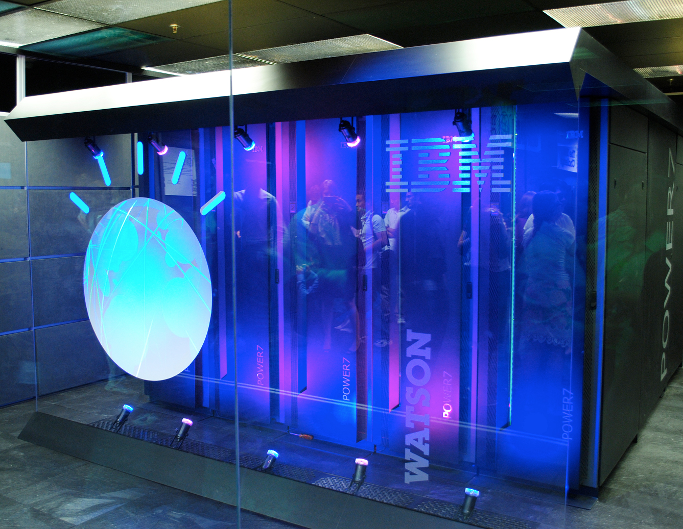
Since the enthusiasm for AI in healthcare brought on by IBM’s Watson, many questions on bias and discrimination in algorithms have emerged. Photo: Wikimedia.

Ethical algorithms in health: a technological and societal challenge

The possibilities offered by algorithms and artificial intelligence in the healthcare field raise many questions. What risks do they pose? How can we ensure that they have a positive impact on the patient as an individual? What safeguards can be put in place to ensure that the values of our healthcare system are respected?


A few years ago, Watson, IBM’s supercomputer, turned to the health sector and particularly oncology. It has paved the way for hundreds of digital solutions, ranging from algorithms for analyzing radiology images to more complex programs designed to help physicians in their treatment decisions. Specialists agree that these tools will spark a revolution in medicine, but there are also some legitimate concerns. The CNIL, in its report on the ethical issues surrounding algorithms and artificial intelligence, stated that they “may cause bias, discrimination, and even forms of exclusion”.

In the field of bioethics, four basic principles were announced in 1978: justice, autonomy, beneficence and non-maleficence. These principles guide research on the ethical questions raised by new applications for digital technology. Christine Balagué, holder of the Smart Objects and Social Networks chair at the Institut Mines-Télécom Business School, highlights a pitfall, however: “the issue of ethics is tied to a culture’s values. China and France for example have not made the same choices in terms of individual freedom and privacy”. Regulations on algorithms and artificial intelligence may therefore not be universal.

However, we are currently living in a global system where there is no secure barrier to the dissemination of IT programs. The report made by the CCNE and the CERNA on digital technology and health suggests that the legislation imposed in France should not be so stringent as to restrict French research. This would come with the risk of pushing businesses in the healthcare sector towards digital solutions developed by other countries, with even less controlled safety and ethics criteria.

Bias, value judgments and discrimination

While some see algorithms as flawless, objective tools, Christine Balagué, who is also a member of CERNA and the DATAIA Institute highlights their weaknesses: “the relevance of the results of an algorithm depends on the information it receives in its learning process, the way it works, and the settings used”. Bias may be introduced at any of these stages.

Firstly, in the learning data: there may be an issue of representation, like for pharmacological studies, which are usually carried out on 20-40-year-old Caucasian men. The results establish the effectiveness and tolerance of the medicine for this population, but are not necessarily applicable to women, the elderly, etc. There may also be an issue of data quality: their precision and reliability are not necessarily consistent depending on the source.

Data processing, the “code”, also contains elements which are not neutral and may reproduce value judgments or discriminations made by their designers. “The developers do not necessarily have bad intentions, but they receive no training in these matters, and do not think of the implications of some of the choices they make in writing programs” explains Grazia Cecere, economics researcher at the Institut Mines-Télécom Business School.

Read on I’MTech: Ethics, an overlooked aspect of algorithms?

In the field of medical imaging, for example, determining an area may be a subject of contention: A medical expert will tend to want to classify uncertain images as “positive” so as to avoid missing a potential anomaly which could be cancer, which therefore increases the number of false-positives. On the contrary, a researcher will tend to maximize the relevance of their tool in favor of false-negatives. They do not have the same objectives, and the way data are processed will reflect this value judgment.

Security, loyalty and opacity

The security of medical databases is a hotly-debated subject, with the risk that algorithms may re-identify data made anonymous and may be used for malevolent or discriminatory purposes (by employers, insurance companies, etc.). But the security of health data also relies on individual awareness. “People do not necessarily realize that they are revealing critical information in their publications on social networks, or in their Google searches on an illness, a weight problem, etc.”, says Grazia Cecere.

Applications labeled for “health” purposes are often intrusive and gather data which could be sold on to potentially malevolent third parties. But the data collected will also be used for categorization by Google or Facebook algorithms. Indeed, the main purpose of these companies is not to provide objective, representative information, but rather to make profit. In order to maintain their audience, they need to show that audience what it wants to see.

The issue here is in the fairness of algorithms, as called for in France in 2016 in the law for a digital republic. “A number of studies have shown that there is discrimination in the type of results or content presented by algorithms, which effectively restricts issues to a particular social circle or a way of thinking. Anti-vaccination supporters, for example, will see a lot more publications in their favor” explains Grazia Cecere. These mechanisms are problematic, as they get in the way of public health and prevention messages, and the most at-risk populations are the most likely to miss out.

The opaque nature of deep learning algorithms is also an issue for debate and regulation. “Researchers have created a model for the spread of a virus such as Ebola in Africa. It appears to be effective. But does this mean that we can deactivate WHO surveillance networks, made up of local health professionals and epidemiologists who come at a significant cost, when no-one is able to explain the predictions of the model?” asks Christine Balagué.

Researchers from both hard sciences and social sciences and humanities are looking at how to make these technologies responsible. The goal is to be able to directly incorporate a program which will check that the algorithm is not corrupt and will respect the principles of bioethics. A sort of “responsible by design” technology, inspired by Asimov’s three laws of robotics.


Article initially written in French by Sarah Balfagon, for I’MTech.

Electroencephalogram: a brain imaging technique that is efficient but limited in terms of spatial resolution.

Technology that decrypts the way our brain works

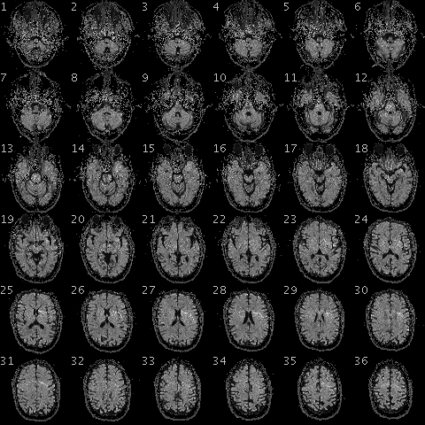
Different techniques are used to study of the functioning of our brain, including electroencephalography, magnetoencephalography, functional MRI and spectroscopy. The signals are processed and interpreted to analyze the cognitive processes in question. EEG and MRI are the two most commonly used techniques in cognitive science. Their performances offer hope and but also concern. What is the current state of affairs of brain function analysis and what are its limits?


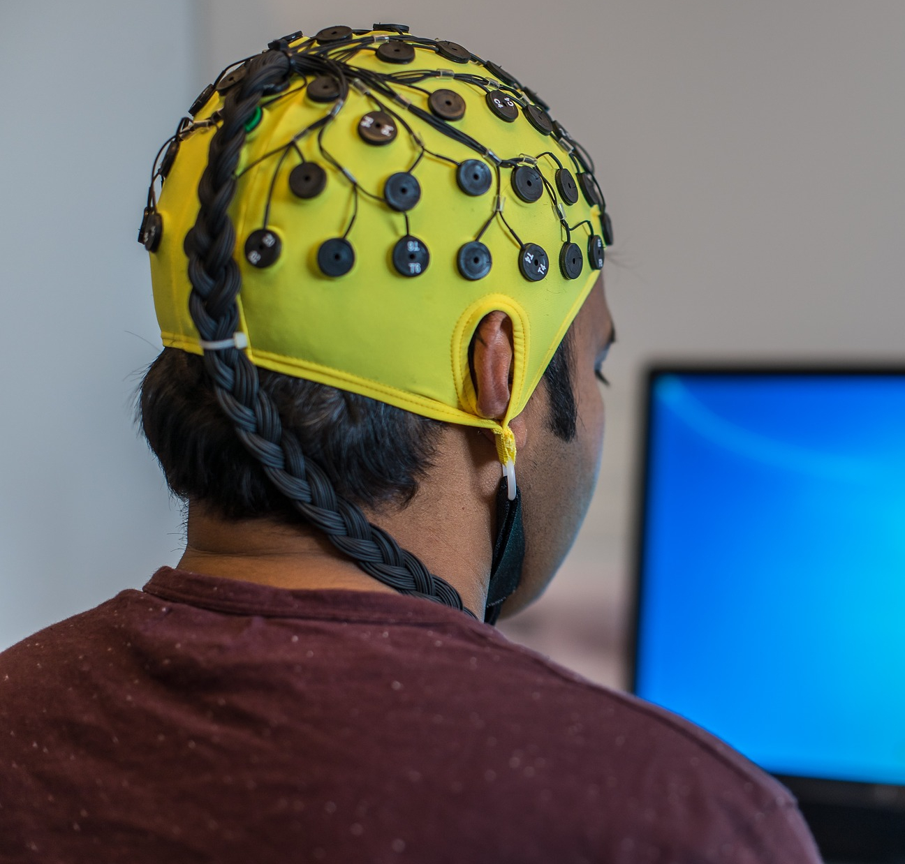
Nesma Houmani is a specialist in electroencephalography (EEG) signal analysis and processing at Télécom SudParis. Neuron activity in the brain generates electrical changes which can be detected on the scalp. These are recorded using a cap fitted with strategically-placed electrodes. The advantages of EEG are that it is not costly, easily accessible and noninvasive for the subjects being studied. However, it generates a complex signal composed of oscillations associated with new baseline brain activity when the patient is awake and at rest, punctual signals linked to activations generated by the test and variable background noise caused, notably, by involuntary movements by the subject.

The level of noise depends, among other things, on the type of electrodes used, whether dry or with gel. While the latter reduces the detection of signals not emitted by brain activity, they take longer to place, may cause allergic reactions and require the patient to thoroughly wash with shampoo after the examination, making it more complicated to carry out these tests outside hospitals. Dry electrodes are being introduced in hospitals, but the signals recorded have a high level of noise.

The researcher at Télécom SudParis uses machine learning and artificial intelligence algorithms to extract EEG markers. “I use information theory combined with statistical learning methods to process EEG time series of a few milliseconds.” Information theory supposes that signals with higher entropy contain more information. In other words, when the probability of an event occurring is low, the signal contains more information and is therefore more likely to be relevant. Nesma Houmani’s work allows the removal of parasite signals from the trace and a more accurate interpretation of the EEG data recorded.

A study published in 2015 showed that this technique allowed better definition of the EEG signal in the detection of Alzheimer’s disease. Statistical modeling allows consideration of the interaction between the different areas of the brain over time. As part of her research on visual attention, Nesma Houmani uses EEG combined with an eye tracking device to determine how a subject engages in and withdraws from a task: “The participants must observe images on a screen and carry out different actions according to the image shown. A camera is used to identify the point of gaze, allowing us to reconstitute eye movements,” she explains. Other teams use EEG for emotional state discrimination or for understanding decision-making mechanisms.

EEG provides useful data because it has a temporal resolution of a few milliseconds. It is often used in applications for brain-machine interfaces, allowing a person’s brain activity to be observed in real time with just a few seconds’ delay. “However, EEG is limited in terms of spatial resolution,” explains Nesma Houmani. This is because the electrodes are, in a sense, placed on the scalp in two dimensions, whereas the folds in the cortex are three-dimensional and activity may come from areas that are further below the surface. In addition, each electrode measures the sum of synchronous activity for a group of neurons.

The most popular tool of the moment: fMRI

Conversely, functional MRI (fMRI) has excellent spatial resolution but poor temporal resolution. It has been used a lot in recent scientific studies but is costly and access is limited by the number of devices available. Moreover, the level of noise it produces when in operation and the subject’s position lying down in a tube can be stressful for participants. Brain activity is reconstituted in real time by detecting a magnetic signal linked to the amount of blood transferred by micro-vessels at a given moment, which is visualized over 3D anatomical planes. Although activations can be accurately situated, hemodynamic variations occur a few seconds after the stimulus, which explains why the temporal resolution is lower than that of EEG.

fMRI produces section images of the brain with good spatial resolution but poor temporal resolution.

fMRI produces section images of the brain with good spatial resolution but poor temporal resolution.


Nicolas Farrugia has carried out several studies with fMRI and music. He is currently working on applications for machine learning and artificial intelligence in neuroscience at IMT Atlantique. “Two main paradigms are being studied in neuroscience: coding and decoding. The first aims to predict brain activity triggered by a stimulus, while the second aims to identify the stimulus from the activity,” the researcher explains. A study published in 2017 showed the possibilities of fMRI associated with artificial intelligence in decoding. Researchers asked subjects to watch videos in an MRI scanner for several hours. A model was then developed using machine learning, which was able to reconstruct a low-definition image of what the participant saw based on the signals recorded in their visual cortex. fMRI is a particularly interesting technique for studying cognitive mechanisms, and many researchers consider it the key to understanding the human brain, but it nevertheless has its limits.

Reproducibility problems

Research protocol changed recently. Nicolas Farrugia explains: “The majority of publications in cognitive neuroscience use simple statistical models based on functional MRI contrasts by subtracting the activations recorded in the brain for two experimental conditions A and B, such as reading versus rest.” But several problems have led researchers to modify this approach. “Neuroscience is facing a major reproducibility challenge,” admits Nicolas Farrugia. Different limitations have been identified in publications, such as a small workforce, a high level of noise and a separate analysis for each part of the brain, not to mention any interactions or the relative intensity of activation in each area.

These reproducibility problems are leading researchers to change methods, from an inference technique in which all available data is used to obtain a model that cannot be generalized, to a prediction technique in which the model learns from part of the data and is then tested on the rest.” This approach, which is the basis for machine learning, allows the model’s relevance to be checked in comparison with the actual reality. “Thanks to artificial intelligence, we are seeing the development of computational calculation methods which were not possible with standard statistics. In time, this will allow researchers to predict what type of image or what piece of music the person is thinking of based on their brain activity.

Unfortunately, there are also reproducibility problems in signal processing with machine learning. The technique, which is based on artificial neural networks, is currently the most popular because it is very effective in multiple applications, but it requires adjusting hundreds of thousands of parameters using optimization methods. Researchers tend to adjust the parameters of the developed model when they evaluate it and repeat it on the same data, thus distorting the generalization of results. The use of machine learning also leads to another problem for signal detection and analysis: the ability to interpret the results. Knowledge of deep learning mechanisms is currently very limited and is a field of research in its own right, so our understanding of how human neurons function could in fact come from our understanding of how deep artificial neurons function. A strange sort of mise en abyme!


Article written by Sarah Balfagon, for I’MTech.


More on this topic:

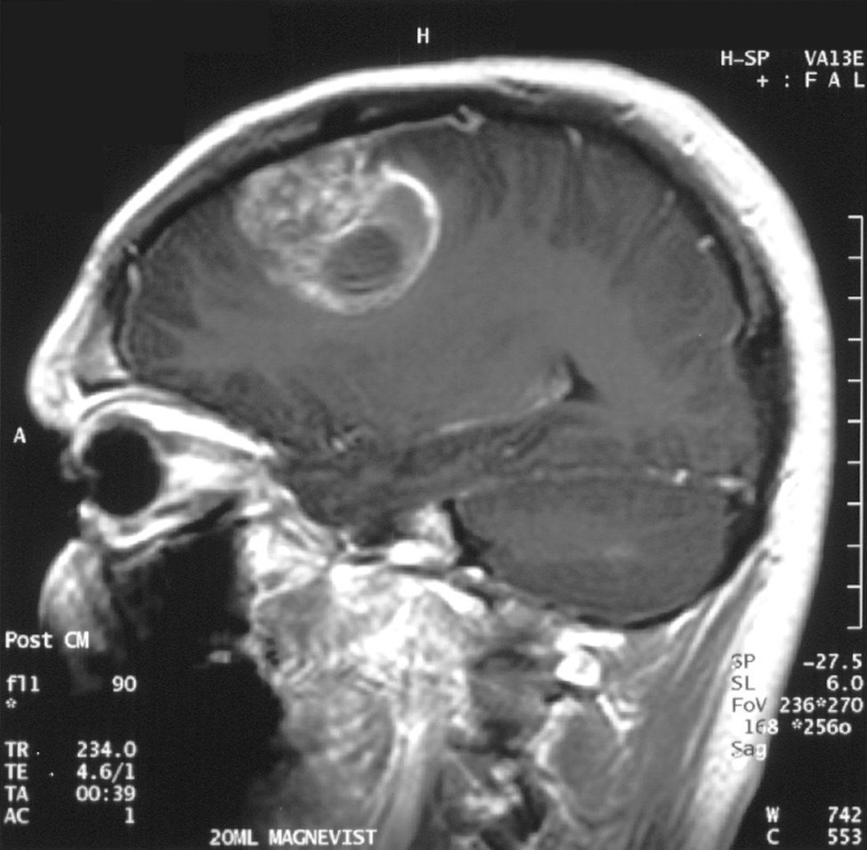
Glioblastoma is a type of brain tumor. It remains difficult to treat. Image: Christaras A / Wikimedia.

Glioblastoma: electric treatment?

At Mines Saint-Étienne, the ATPulseGliome project is looking into a new form of cancer treatment. This therapeutic approach is aimed at fighting glioblastoma, an especially aggressive form of brain cancer, using electrical stimulation. It could eventually increase the life expectancy of glioblastoma patients in comparison with chemotherapy and radiotherapy treatment. 


Glioblastoma is a rare form of brain cancer. Of the 400,000 new cases of cancer recorded each year in France, it affects 2,400 people, or 0.6%. Unfortunately, it is also one of the more severe forms, with the life expectancy of glioblastoma patients at between 12 to 15 months with treatment. To improve the survival chances of those affected, Mines Saint-Étienne is leading the ATPulseGliome project, in collaboration with the University of Limoges and with funding from the EDF Foundation. The team of researchers, led by Rodney O’Connor, is testing an approach using electric fields to find new types of treatment.

The glial cells are located around the neurons in our brain. These are the cells affected by glioblastoma. “One particular type of glial cell is affected: the astrocytes” says Hermanus Ruigrok, a cellular biologist at Mines Saint-Étienne and researcher on the ATPulseGliome project. The normal role of astrocytes is to provide nutrients to neurons, repair brain lesions, and ensure separation between the nervous system and blood circulation system. Like all cells, astrocytes regenerate by division. Glioblastoma survive when the astrocytes behave abnormally, dividing in an uncontrollable manner.

Targeting glioblastoma without affecting healthy cells

ATPulseGliome is looking into a form of treatment based on electrical stimulation of cancer cells. “Healthy astrocytes are not sensitive to electricity, but glioblastoma cells are” explains Hermanus Ruigrok. This difference is the foundation of the treatment strategy, which will target the cancer cells only, not the astrocytes and other healthy cells. Glioblastoma cancer cells have a larger amount of proteins in their membrane which are sensitive to electricity.

These proteins act as doors, letting ions in and out, thus enabling communication between the cell and the outside environment. This door malfunctions under the effect of electrical stimulus. An unusually high number of ions then enter the cancer cell, causing harmful effects. “This strategy will allow us to destroy only the cancer cells, and not the healthy astrocytes, which are not sensitive to the electrical stimulus” Hermanus Ruigrok highlights.

Glioblastoma cells, marked with fluorescence.

Glioblastoma cells, marked with fluorescence.


It is still much too early to trial this technique on patients. The ATPulseGliome team is working on glioblastoma cell lines. Initially, these cells come from a patient with this form of cancer, but they are cultivated, reproduced and isolated in in vitro experiments. By eliminating the complex molecular interactions present in a real patient, this first step helps to clarify the scientific objectives and test the feasibility of in vivo tests. During this phase, researchers will look at the different types of electrodes to be used for sending an electrical field, determine the characteristics of the electrical signal required in stimulating cells, and measure the initial responses of glioblastoma to the electrical impulses.

To complete these steps, the team at Mines Saint-Étienne is working with Institut des neurosciences de la Timone in Marseille. “We want to take on as many specialists as possible, as the project requires a range of skills: biologists, electronics engineers, neurologists, surgeons, etc.”, Hermanus Ruigrok explains. Although it is a lengthy procedure, this multidisciplinary approach could increase the life expectancy of patients. “We can’t say in advance how much more effective this electrical field approach will be compared with the chemotherapy and radiotherapy currently used”, the researcher explains. “Although it may be difficult to fully cure this cancer, being able to monitor and limit its development in order to significantly increase the life expectancy of those affected by glioblastoma would be a form of satisfaction.”


Hospitals

Improving organization in hospitals through digital simulation

How can we improve emergency room wait times, the way scheduled hospitalizations are managed and cope with unexpected surges of patients? Vincent Augusto, a researcher in healthcare systems engineering at Mines Saint-Étienne is working to find solutions to these problems. He is developing programs based on digital simulation, aimed at optimizing influxes of patients and waiting times at the hospital, especially in emergency care facilities.


Chronic emergency department saturation and unacceptable wait times for receiving care are regularly listed among areas in need of improvement. Several of these areas have been studied: taking preventive action beforehand to reduce influxes of patients, organization within emergency departments, managing hospitalizations in advance. Vincent Augusto and his team from the MedTechDesign living lab at the engineering and healthcare center at Mines Saint-Étienne have developed models that contribute to these last two areas by providing valuable information. “We worked on successive projects with hospitals to develop programs using digital simulation. The principle is that any system can potentially be monitored and reproduced based on the data it generates; being able to process this data in real time would help to optimize resources. Unfortunately, major inequalities exist in terms of computerization from one hospital to another.

Vincent Augusto is specialized in modeling, analyzing and managing inflows of patients in hospitals. “At the hospital in Firminy, we modeled unforeseen arrivals in the emergency department to get a better idea of the number of beds required and to improve planning for scheduled patients.” The departments schedule hospitalizations for patients needing diagnostic scans or treatment. However, since it is difficult to predict the number of available places in advance, scheduled hospitalizations must sometimes be canceled at the last minute, forcing patients to wait longer to receive care. On the other hand, the shortage of beds leads to overcrowded emergency services. Improving the management of the internal and external flow of patients in hospitals is therefore of utmost importance.

A modular digital twin

At the university hospital (CHU) in Saint-Étienne, the team developed a digital twin for the emergency department. This twin helped assess the different measures that could be implemented to improve emergency operations. Vincent Augusto explains how this was developed: “First, there is an on-site observation phase. We collect data using existing software. Next, there is a development phase in which we seek to understand and model the flow of patients in the department and create an initial model on paper that is confirmed by the department staff. We can then create a digital assessment model that reproduces the way the emergency department operates, which then undergoes a validation phase.”

The researchers use the department’s activities from the previous year to accomplish this. They enter the data into the system and check if the indicators predicted by the model match those recorded at the time. This approach involves three different components: the first analyzes the patient care circuit, the second analyzes human resources based on type of activity and the third focuses on the organization and interdependence of the resources. “Once this model has been validated, we can use the modular system to test different scenarios: we can alter the human resources, simulate the arrival of an inflow of patients, reduce the wait time for taking further tests—such as scans—or the time required to transfer a patient to a hospital ward,” the researcher explains.

The first measure tested was to divide emergencies into three groups: serious emergencies (road accidents, respiratory problems, etc.), functional emergencies (sprains, wounds requiring stitches, etc.) and fast functional emergencies (requiring care that can be quickly provided). Upon entering, the patients are directed to one of these three groups led by different teams. According to Vincent Augusto and the system users, “this makes it possible to clearly assess the savings in terms of time and costs that are related to organizational changes or an increase in human resources, before any real changes are made. This is a big plus for the departments, since organizational changes can be very time-consuming and costly and sometimes have only a small impact.”

The real impact the organizational measures would have on emergency department operations was assessed and made it possible to continue work on another potential area for improvement: the creation of a psychiatric section within the emergency department, with beds reserved for these patients. To help draw up the plans for the future emergency services, the team from Mines Saint-Étienne is developing a virtual reality interface to directly and realistically view flows of patients more easily than the indicators and charts generated by the digital simulation system. The goal is to optimize the patient circuit within the department and the medical care they receive.

Improving hospitals’ resilience in unexpected events

This method also offers management support for crisis situations involving a massive influx of patients to the emergency department in the event of disasters, attacks or epidemics. “The system was developed to manage, in addition to the usual flow of patients, an exceptional yet predictable arrival of patients,” the researcher explains. It is therefore useful in voltage plans: exceptional situations that push the system beyond its capacity. In these cases, the department must face a critical situation of responding to hospital emergencies that can lead to a French emergency “white plan” being declared, in which non-priority activities are cancelled.

To accomplish this, the program is updated in real time via a direct connection to the hospital’s computer systems. It can therefore determine the exact state of the department at any time. By entering a varying number of patients with specific pathologies in a given situation (flu-related respiratory difficulties, gunshot wounds, etc.), the simulation can determine the most effective measures to take. This is what the engineers call an operational tool. “In the short and medium term, the departments now have a tool that can help them optimize their response to the problems they face and improve the care patients receive,” concludes Vincent Augusto.

Original article in French written by Sarah Balfagon, for I’MTech.