Temporary tattoos for brain exploration

A team of bioelectronics researchers at Mines Saint-Étienne has developed a new type of electroencephalogram electrode using a temporary tattoo technique. As effective as traditional electrodes, but much more comfortable, they can provide extended recordings of brain activity over several days. 


The famous decalcomania transfer technique – made popular in France by the Malabar chewing gum brand in the 1970s – has recently come back into fashion with temporary tattoos. But it does not serve solely to provide fun for people all ages. A new use has been developed with the invention of temporary tattoo electrodes (TTE) designed to record electrophysiological signals.

Originally developed to pick up heart (electrocardiogram, ECG) and muscle signals (electromyogram, EMG), the technique has been refined to reach the holy Grail of bioelectronics: the brain. “Electroencephalographic signals (EEG) are the hardest to record since their amplitudes are lower and there is more background noise, so it was a real challenge for us to create flexible epidermal electronic devices that are as effective as standard electrodes,” explains Esma Ismailova a bioelectronics researcher at Mines Saint-Étienne.

From Pontedera to Saint-Étienne

The process for printing tattoo electrodes was developed by an Italian team, led by Francisco Greco, at the Italian Institute of Technology in Pontedera. The next step for preclinical application was carried out at the Saint-Etienne laboratory. Laura Ferrari, a PhD student who worked on TTE with Francisco Greco for her thesis, chose to carry out postdoctoral research with Esma Ismailova in light of her experience in the field of wearable connected electronics. In 2015, the Mines Saint-Étienne team had developed a connected textile, derived from the technique used to print on kimonos, intended to record an electrocardiogram on a moving person, with fewer artifacts than traditional ECG electrodes.

The sensors of the tattoo electrodes, like the textile electrodes, are composed of semi-conductive polymers. These organic compounds, which were the topic of the 2000 Nobel prize in chemistry, act as transistors and offer new possibilities in the field of surface electronics.  The conductive polymer used is called PEDOT:PSS. It is mixed with ink and projected on a paper sold commercially for temporary tattoos, using a regular inkjet printer. The back layer is removed at the time of application. A simple wet sponge dissolves the soluble layer composed of cellulose, and the tattoo is transferred to the skin. The materials and techniques used in the microfabrication process for TTEs make it suitable for large-scale, low-cost production.

Esma Ismailova and her team worked extensively on the assembly and interconnection between the electrodes and electronic signal recording devices. An extension ending in a plastic clip was manufactured through 3D printing and integrated in the decalcomania. The clip makes it possible to attach a wire to the tattoo: “We had to solve the problem of transmitting the signal to transfer the data. Our goal is now to develop embedded electronics along with the electrodes, a microfabricated, laminated board on the patch to collect and memorize information, or transmit it through a mobile phone,” says the Saint- Étienne researcher.

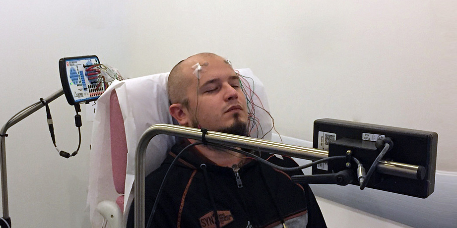
a: multi-layer structure of a TTE allowing for the transfer of the top film on which the electrode is printed b: exploded view of a TTE with integrated flat connection c: TTE transferred to the scalp in the occipital region d: close-up of a TTE 12h after application with hair regrowth

Electrodes that are more comfortable for patients…

The dry electrodes composed of a one-micron thick polymer film conform perfectly to the surface of the skin due to their flexibility. This interface makes it possible to dispense with the gel which is necessary for traditional electrodes, which dries out after a few hours, making the electrodes inoperative.  The transfer must be done on shaved skin, but a study has shown that hair regrowth through the film does not stop them from being effective. This means that they can be used for 2 to 3 days, provided they do not get wet, since the principle of temporary tattoos is that they are broken down by washing with soap and water.  Research is currently underway  to replace the regular transfer layer by a more resistant, water-repellent material, which would extend their lifetimes.

For Esma Ismailova, this technology is a huge step forward, for both the field of clinical research and patient care: “These new flexible, stretchable, very thin electrodes are ergonomic, conformable, virtually imperceptible, and are therefore much more acceptable for patients, particularly children and elderly people, for whom certain exams can be stressful.”  Indeed, to perform an EEG, patients must normally wear a headset that attaches below the chin, composed of electrodes on which the technician applies gel.

… and more effective for doctors

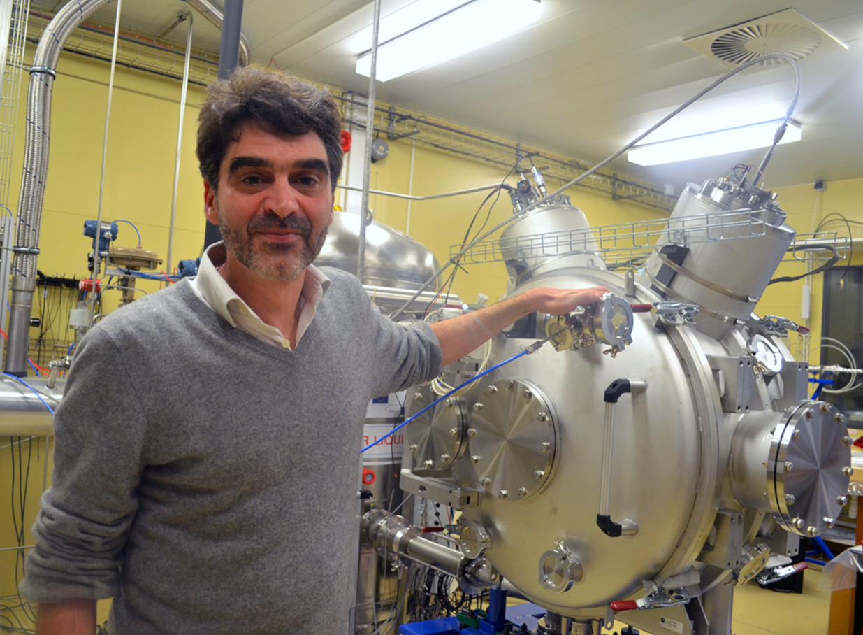
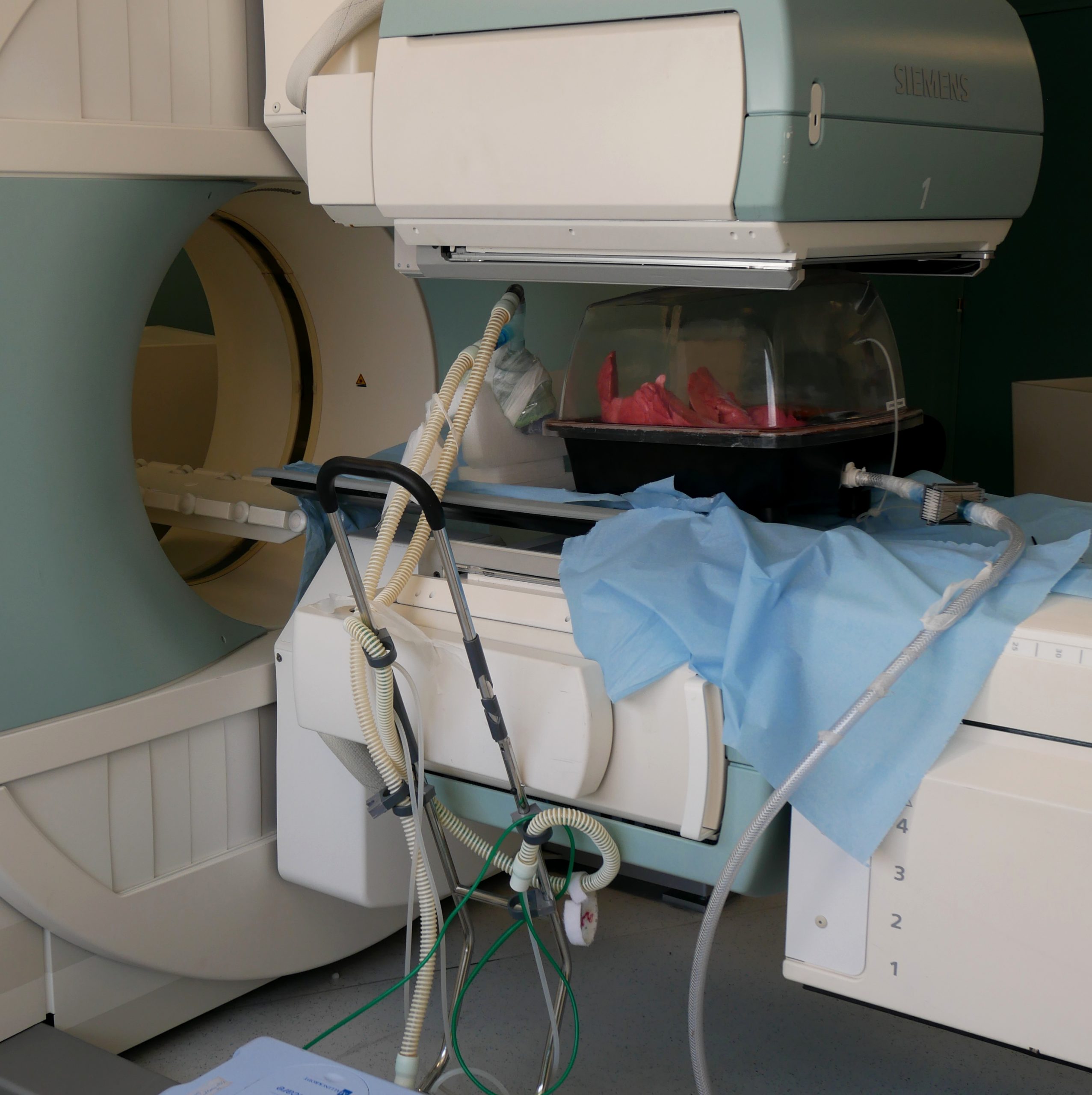
Another advantage of these temporary tattoo electrodes is their compatibility with  magnetoencephalography (MEG). Since they are composed entirely of organic materials and therefore do not contain any metal, they do not disturb the magnetic field generated by the device and do not create artifacts, so they can be used to perform EEGs coupled with MEGs. These two techniques for exploring neuronal activity are complementary and refine information about the  starting point of epileptic seizures,  the review of systems for certain tumors before their ablation, and neurodegenerative diseases.

The clinical assessment of TTE in the field of neurophysiology was carried out in collaboration with Jean-Michel Badier from the Institut de Neurosciences des Systèmes at the University of Aix-Marseille. This study was recently published in the journal Nature, and confirmed that their performance was similar to traditional electrodes for standard EEG, and superior for MEG, since they do not produce any shadow areas.

“We’ve done a proof of concept, now we’re trying to develop a device that can be used at home. We plan to do a study with epileptic or autistic children, for whom comfort and acceptability are very important,” explains Esma Ismailova. These tattoo electrodes – like other connected technology –will generate a great amount of data. For the researcher, “it’s essential to collaborate with researchers who can process this data using specialized algorithms. It’s a new era for smart wearables designed for personalized, preventive medicine, in particular through the early detection of abnormalities.”


Sarah Balfagon

masks

Protective masks: towards widespread reuse?

How can protective masks be recycled and reused without risking safety? Scientists, medical practitioners and manufacturers have teamed up to explore different treatment methods. As part of this consortium, IMT Atlantique researchers are studying the impact of decontamination processes on mask performance.


Surgical and FFP2 masks are intended for single use. Thrown away after just a few hours of use, they are designed to protect the wearer from inhaling infectious agents spread through the air. The question of recycling these masks has not been raised before, but high demand for masks to protect healthcare workers and the general public has been a game-changer. To help find a solution to the current shortage, an interdisciplinary consortium bringing together nearly 25 laboratories and manufacturers throughout France was created in early March, led by professor Philippe Cinquin from Grenoble University Hospital, the CNRS and the CEA. Its goal is to find a treatment process that makes it possible to reuse masks.

Currently, various decontamination methods recognized for both their virucidal and bactericidal effects are being explored: among others, gamma or beta irradiation, thermal decontamination with steam at 121 °C; an ethylene oxide treatment, and wet or dry heating methods at 70°C or higher. These methods must able to reduce the bioburden of protective masks, without reducing filtration efficiency or breathability.

At IMT Atlantique, which is a member of the research consortium, Laurence Le Coq and her colleagues Aurélie Joubert and Yves Andrès are working mainly on this second aspect of the project.  The researchers are drawing on their research on filtration applied to air treatment — for industrial waste applications, for example, or indoor air treatment in ventilation networks. The team has been able to quickly shift its focus and adapt its expertise to work on recycling used masks. “The contribution and dedication of researchers and technical staff, who were called on to respond to an urgent need to develop technical solutions and establish experimental conditions, has been instrumental,” says Laurence Le Coq.

By mid-March, the scientists had set up an initial test bed to closely reflect the AFNOR standards for masks in order to test their performance following decontamination. “If the masks are normally intended for single use, it’s also because they first undergo a treatment process which gives them a certain level of efficiency, as well as their mechanical strength and specific shape. When they are decontaminated, part of this pre-treatment is removed, depending on the type of decontamination and its conditions. What’s more, depending on how a mask has been put on, worn and taken off, it may be damaged and its structure could be altered,” explains the researcher.

Preliminary findings    

So, how can effective decontamination be combined with a sufficient level of protection?  “We compare how performance is maintained between new treated masks and used treated masks. More precisely, we measure the changes brought about by decontamination treatments, in particular their level of breathability and their filtration efficiency for particles with a diameter ranging from 0.3 to 3 µm, since the virus is spread by microdroplets,” explains Laurence Le Coq.

After ruling out certain methods, the scientists were able to determine favorable treatment conditions for decontaminating the masks without having too much of an effect on their inherent qualities. “Dry heat treatments, for example, are promising but we can’t move forward for the time being. Certain findings are encouraging following irradiation or washing at 95°, but only for surgical masks. For now, our findings do not allow us to converge on a single treatment, a single protocol. And most importantly, there is a huge difference between what we do in good laboratory conditions and what could be done on a greater quantity of masks in a hospital environment, or at home,” says Laurence Le Coq.

The researchers are currently trying to clarify and confirm these preliminary findings. Their goal is now to quickly establish treatment conditions that are effective for all surgical and FFP2 masks, regardless of the manufacturer.

Is widespread mask recycling possible?

The majority of masks provided for the “general public” during the lockdown are reusable after being washed at 60°C for 30 minutes. This is not yet the case for professional masks.

Lockdown measures have been accompanied by efforts to raise public awareness about the importance of wearing masks. In French departments classified as red due to a high number of cases, masks intended for the “general public” are mandatory on public transportation and in high schools. These fabric masks are less effective than professional protective masks, but they are easily reusable and can be washed at least five times. “What is lost in effectiveness is made up for by widespread mask-wearing and ease of use,” says Laurence Le Coq.

For the researcher, this unprecedented research project could also be an opportunity to consider recycling protective masks in the long term, even when there is not a shortage. “Is it really appropriate to have single-use masks if at some point we are required to use them to a greater extent, or even on a daily basis? How should the environmental costs of this medical waste be weighed? Of course, what happens next will depend on the treatment we’re able to develop.”


By Anne-Sophie Boutaud

Reducing the duration of mechanical ventilation with a statistical theory

A team of researchers from IMT Atlantique has developed an algorithm that can automatically detect anomalies in mechanical ventilation by using a new statistical theory. The goal is to improve synchronization between the patient and ventilator, thus reducing the duration of mechanical ventilation and consequently shortening hospital stays. This issue is especially crucial for hospitals under pressure due to numerous patients on respirators as a result of the Covid-19 pandemic.


Dominique Pastor never imagined that the new theoretical approach in statistics he was working on would be used to help doctors provide better care for patients on mechanical ventilation (MV). The researcher in statistics specializes in signal processing, specifically anomaly detection. His work usually focuses on processing radar signals or speech signals. It wasn’t until he met Erwan L’Her, head of emergencies at La Cavale Blanche Hospital in Brest, that he began focusing the application of his theory, called Random Distortion Testing, on mechanical ventilation. The doctor shared a little known problem with the researcher, which would become a source of inspiration: a mismatch that often exists between patients’ efforts while undergoing MV and the respirator’s output.

Signal anomalies with serious consequences

Respirators–or ventilators–feature a device enabling them to supply pressurized air when they recognize demand from the patient. In other words, the patient is the one to initiate a cycle. Many adjustable parameters are used to best respond to an individual’s specific needs, which change as the illness progresses. These include inspiratory flow rate and number of cycles per minute. Standard settings are used at the start of MV and then modified based on flow rate/ pressure curves–the famous signal processed by the Curvex algorithm, which resulted from collaboration between Dominique Pastor and Erwan L’Her.

Patient-ventilator asynchronies are defined as time lags between the patient’s inspiration and the ventilator’s flow rate. For example, the device cannot detect a patient’s demand for air because the trigger threshold level is set too high. This leads to ineffective inspiratory effort. It can also lead to double triggering when the ventilator generates two cycles for one patient inspiratory effort. The patient may also not have time to completely empty their lungs before the respirator begins a new cycle, leading to dynamic hyperinflation of the lungs, also known as intrinsic PEEP (positive end-expiratory pressure).

Effort inspiratoire inefficace : la demande du patient n’aboutit pas à une insufflation

Example of ineffective inspiratory effort: patient demand does not result in insufflation.


Double déclenchement : un seul effort inspiratoire aboutit à deux insufflations rapprochées

Example of double triggering: a single inspiratory effort results in two ventilator insufflations within a short time span.


PEP intrinsèque : l’insufflation suivante survient alors que le débit n’est pas nul à la fin de l’expiration

Example of positive end expiratory pressure: the next ventilator insufflation occurs before the flow has returned to zero at the end of expiration.


These patient-ventilator anomalies are believed to be very common in clinical practice. They have serious consequences, ranging from patient discomfort to increased respiratory efforts that can lead to invasive ventilation–intubation. They involve an increase in the duration of mechanical ventilation, with an increase in weaning failure (end of MV) and therefore longer hospital stays.

However, the number of patients in need of mechanical ventilation has skyrocketed with the Covid-19 pandemic, while the number of health care workers, respirators and beds has only moderately increased, which at times gives rise to difficult ethical choices. A reduction in the duration of ventilation would therefore be a significant advantage, both for the current situation and in general, since respiratory diseases are becoming increasingly common, especially with the aging of the population.

A statistical model that adapts to various signals

Patient-ventilator asynchronies result in visible anomalies in air flow rate and pressure curves. These curves model the series of inspiratory phases, when pressure increases and expiratory phases, when it decreases, with inversion of the air flow. Control monitors for most next-generation devices display these flow rate and pressure curves. The anomalies are visible to the naked eye, but this requires regular monitoring of the curves, and a doctor to be present who can adjust the ventilator settings. Dominique Pastor and Erwan L’Her had a common objective: develop an algorithm that would detect certain anomalies automatically. Their work was patented under the name Curvex in 2013.

The detection of an anomaly represents a major deviation from the usual form for a signal. We chose an approach called supervised learning by mathematical modeling,” Dominique Pastor explains. One characteristic of his Random Distorsion Testing theory is that it makes it possible to detect signal anomalies with very little prior knowledge. “Often, the signal to be processed is not well known, as in the case of MV, since each patient has unique characteristics, and it is difficult to obtain a large quantity of medical data. The usual statistical theories have difficulty taking into account a high degree of uncertainty in the signal. Our model, on the other hand, is generic and flexible enough to handle a wide range of situations.” 

Dominique Pastor first worked with intrinsic PEEP detection algorithms with PhD student Quang-Thang Nguyen, who helped to find solutions. “The algorithm is a flow rate signal segmentation method used to identify the various breathing phases and calculate models for detecting anomalies. We introduced an adjustable setting (tolerance) to define the deviation from the model used to determine whether it is an anomaly,” Dominique Pastor explains. According to the researcher from IMT Atlantique, this tolerance is a valuable asset. It can be adjusted by the user, based on their needs, to alter the sensitivity and specificity.

The Curvex platform not only processes flow data from ventilators, but also a wide range of physiological signals (electrocardiogram, electroencephalogram). A ventilation simulator was included, with settings that can be adjusted in real-time, in order to test the algorithms and perform demonstrations. By modifying certain pulmonary parameters (compliance, airway resistance, etc.) and background noise levels, different signal anomalies (intrinsic PEEP, ineffective inspiratory effort, etc.) appear randomly. The algorithm detects and characterizes them. “In terms of methodology, it is important to have statistical signals that we can control in order to make sure it is working and then move on to real signals,” Dominique Pastor explains.

The next step is to create a proof of concept (POC) by developing electronics to detect anomalies in ventilatory signals, to be installed in emergency and intensive care units and used by health care providers. The goal is to provide versatile equipment that could adapt to any ventilator. “The theory has been expanding since 2013, but unfortunately the project has made little progress from a technical perspective due to lack of funding.  We now hope that it will finally materialize, in partnership with a laboratory, or designers of ventilators, for example. I think this a valuable use of our algorithms, both from a scientific and medical perspective,” says Dominique Pastor.

By Sarah Balfagon for I’MTech.

Learn more:

– Mechanical ventilation system monitoring: automatic detection of dynamic hyperinflation and asynchrony. Quang-Thang Nguyen, Dominique Pastor, François Lellouche and Erwan L’Her

Illustration sources:

Curves 1 and 2

Curve 3


STREAM

STREAM: Bone tissue model culture

The STREAM project, which Mikhaël Hadida is working on at Mines Saint-Étienne, aims to develop a controlled, carefully-managed bone tissue culture platform. This system would facilitate observation in order to study the mechanisms involved in bone tissue by reducing the costs and time required for research.  


A culture system for bone tissue models in a controlled environment that allows for simplified observations: this is the aim of the STREAM project. Led by Mikhaël Hadida, a researcher at Mines Saint-Étienne, the STREAM research project (System for Bone Tissue Relevant Environment And Monitoring) aims to provide academic and industrial players with a useful tool for developing simplified, standardized and automated bone tissue models.

This in vitro culture system seeks to provide an innovative alternative to avoid animal testing in bone biology research. It is designed for laboratories, but could also be used to validate a medical system for pharmaceutical compounds or medical devices. This could pave the way for advances in research on osteoporosis, for example, or help make living bone grafts for regenerative medicine applications.

“The structure allows us to control the mechanical parameters of the system,” explains Mikhaël Hadida. It will be possible to control the culture conditions “and measure cell activity by collecting key data in real time.” The ability to control the mechanical environment of this type of culture has never before been reported in scientific literature. This would allow for better reproducibility between experiments, a key issue in research, while lowering costs and reducing the time required for research.

Little-understood mechanisms

One of the problems with current systems is the lack of a controlled environment. If the environment is not homogenous, it is difficult to draw conclusions from the experiments. “In research, we still don’t have a clear understanding of what is involved in mechanical stress on bone tissue,” explains Mikhaël Hadida.

Current systems are generally designed using biomimicry based on demineralized bone.   They remain very close to the human system and therefore imply better performance. “But it’s also a very complex system that is not possible to control or manage,” says the researcher.

There may be other phenomena involved, which in turn could affect the results observed. It is difficult to determine whether the parameters used have a real impact on the culture, or if other mechanisms the researchers are not aware of come into play. New collagen-based structures have also been developed, but they are still based on these highly complex systems. “It’s an attempt to find a shortcut – seeking performance before the foundations of these mechanisms are properly understood,” he adds.

The Bone Stream system

Mechanical stress in bones is not directly related to wear and tear, but bone cells react to mechanical stimuli that can damage bone tissue. Our bones are made up of a solid matrix and pore fluid. Walking or running leads to micro-stimulations – micro-compressions on this fluid – which create “currents” that stimulate the mechanisms involved.

“At the Centre for Biomedical and Healthcare Engineering, David Marchat can make us tailor-made culture scaffolds, which are perforated,” explains Mikhaël Hadida. The system the research team is working on is perfused to reproduce these fluid movements in the culture medium and obtain conditions similar to those of real life. “So we have a good combination in terms of the culture chamber and the culture medium to ensure that all the bone cells have a homogenous scaffold.”

Read more on I’MTech: Bone implants to stimulate bone regeneration

This homogeneity is essential to control the system and understand what impacts the culture, so as to be certain that it is indeed the mechanisms used that are responsible and not another variable the researchers are not aware of. Parameters tested include, for instance, fluid flow speed and what is known as shear stress. If you put pressure on a cube by moving your hand down its side, you apply a force parallel to the surface: this is shear stress.

Direct observation

“This system would also offer a major advantage for making observations,” says Mikhaël Hadida. “Current culture systems are destructive. In order to observe the culture they must be interrupted,” he explains. The culture scaffold must first be taken out of its environment. Then cell markers must be injected and it must be cut into very fine slices so that they can be examined under a microscope. It can also be ground up to analyze the DNA.

If you wish to study the development of your culture, these destructive methods pose a major problem. “If you want to observe your culture on a given day, then two days later, then a week later, your needs increase with all these observations,” he says. This means culture scaffolds must be available for each observation, which requires a significant investment, in terms of time and costs.

“So we wanted to develop a platform that could observe the culture in real time,” says the researcher. Their culture scaffold is installed on the transparent inner face of the culture chamber. This extends into a viewing chamber to monitor its evolution with a microscope without disturbing the culture scaffold. “We also have sensors to study the evolution of cell activity,” he adds. “They monitor changes in the culture medium very closely and therefore keep track of how the cells develop.”

The goal is now to continue developing this platform to bring a culture tool to the market that stands out from those already available. “For example, we’re working on a project with the European Space Agency (ESA) on this topic,” says Mikhaël Hadida. The astronauts on board the International Space Station (ISS) are subjected to specific mechanical stress and present a phenomenon of accelerated aging of bone tissue upon their return. ESA is therefore actively involved in this research in order to better understand this phenomenon and find solutions.


By Tiphaine Claveau, for I’MTech

ERC

Twin ERC grants for research on the aorta

In 2015, the Mines Saint-Étienne engineering and health center was awarded two grants by the European Research Council (ERC). This funding was for two five-year projects on ruptured aortic aneurysms in the Sainbiose laboratory[1]Pierre Badel received a €1.5 million starting grant (young researcher grant), and Stéphane Avril received a €2 million consolidator grant (for putting together a research team). 2020 marks the end of their grants and the related research projects. On this occasion, I’MTech conducted a joint interview with these two researchers to discuss their results and the impact of these ERC grants on their work.


Your two ERC grants were awarded in 2014 and started in 2015, focusing on similar topics: the biomechanics of the aorta in the context of ruptured aneurysms. What were the particularities of each of your projects?

Pierre Badel: The starting point for my project, AArteMIS, was to better explain the resistance of the walls of the aorta. In 2014, we had just developed in vitro tests to study the mechanical strength of this artery. The ERC grant was used to add experiments on the microstructure. In concrete terms, developing protocols to draw on these materials and study the structural properties when the wall breaks.

Stéphane Avril: My Biolochanics project had a degree of overlap with AArteMIS. We had recovered aneurysm tissue from real patients through our partnership with Saint-Étienne University Hospital, and we wanted to characterize the mechanical stresses in these tissues in order to understand how an aneurysm develops and how it ruptures. The two projects were not designed to work together, it is not common to have two ERC grants in the same team. However, the evaluation committees for the starting grant and the consolidator grant applications are different, which meant that the two projects were judged independently and both ended up receiving grants. The connection between the two projects was made afterwards.

Reproduction in vitro d'une dissection aortique pour l'étude de la rupture d'un anévrisme. Ici, l'image est réalisée par tomographie à rayons X sur une artère de lapin.

In vitro reproduction of an aortic dissection for the study of aneurysm rupture. Here, the image is made by X-ray tomography on a rabbit artery.


How did you adapt the research in each of the projects to what was being done in the other?

SA: When we learned that we had been awarded the two scholarships, I redesigned my project. I turned my focus towards the mechanical and biological aspects of the research. Rather than studying the mechanical reasons for aneurysm rupture and their relationship to artery wall structure – which the AArteMIS project was already doing – I focused on early aortic wall changes and their relationship to the environment. For example, the study looked at how blood flows through the aorta and how this affects the development of the aneurysm. We also launched a new protocol in the project to include patients with very small aneurysms. We are still monitoring these patients today, and this gives us a better understanding of the development of the pathology.

PB: For my part, I stayed fairly close to the planned program, i.e. the mechanical study of the material of the artery. The only difference with the original project is that we were able to look further into the structural aspect in the rupture of walls. We had the opportunity to use a new technique: X-ray tomography. This is like a CT scan, but suitable for very small samples. This allowed us to work on each layer of the vessels that make up the wall of the aorta, which have different properties.

These two projects have gone on for five years and will come to an end in a few months. What are the key findings?

PB: For AArteMIS, we now have experience that proves that even if we know the precise thickness of an aneurysm, we cannot predict where it will break. We are very proud of this result because a material will usually break at its thinnest point. However, this approach is wrong. This result helps in the diagnosis of aneurysms by explaining to practitioners that there is more involved than just looking at the thickness of the aortic wall when deciding whether an aneurysm is at risk of rupture.

What about the results of the Biolochanics project?

SA: There are two things I’m very happy about. The first is having finished a scientific article that took 5 years to write. It concerns the development of a method to reconstitute the elasticity map of vessels. It’s a very interesting technique because no one had managed to make an elasticity map of the vessels in the aorta before us. We have filed a patent, and the technique could be used in pharmacological research. The second result is that we have developed a digital model to simulate the accelerated aging of the aorta according to biological parameters. This is a step towards the development of a digital twin of the aorta for patients.

The research conducted under Stéphane Avril’s ERC grant has led to the development of a digital model to simulate the development of an aneurysm (in red on the right) based on biological parameters of the initial artery (left).


Read on I’MTech: A digital twin of the aorta to prevent aneurysm rupture

An ERC grant provides major funding over five years. How do these funds help you to develop a research project in concrete terms?

PB: First of all, an ERC grant means that for a few years we don’t have to waste time looking for money. This is a great comfort for researchers, who constantly have to apply for funding to conduct their work. Specifically for my project, the grant allowed me to recruit three PhD students and three post-docs. A whole team was put together, and that has given us greater research power. In our discipline, there are also many experiments involving expensive tools and equipment. The grant makes it possible to acquire state-of-the-art equipment and to set up the experiments that we wish.

SA: It’s similar for me: we were able to hire nine post-docs for Biolochanics. That’s a considerable size for a research team. The financial comfort also means that you can devote time to scientific resourcing and collaborations. I have been able to spend one to two months each year at Yale University in the United States, where there is also a very good team in specialized biomechanics of the aorta, led by Jay Humphrey.

Read on I’MTech: How Biomechanics can Impact Medicine – Interview with Jay Humphrey

How does being responsible for a project funded by an ERC grant affect your life as a researcher?

SA: There’s a lot of time spent managing and organizing. It’s demanding, but you can see the benefits for the laboratory directly. It is time that is well spent, and that is the main difference from having to spend time looking for funding, where the outcome is more uncertain. It also means a lot of recognition for the work. As researchers, we are solicited more often, we receive invitations that probably would not have come without the ERC grant. In terms of international interaction, it makes a significant difference.

As you approach the end of the projects – the end of December for you Stéphane, and the end of October for you Pierre – how do you envisage the future of your research?

PB: Right now we’re at full throttle! We still have several scientific articles in progress. The project officially ends in the fall, so I’m slowly starting to look for funding again. For example, I have a local project that is about to start up on soft tissue rupture for abdominal wall repair, funded by the Rhône-Alpes Region, Lyon University Hospital, Insa Lyon, and Medtronic. But the next few months will still be very busy with the end of the AArteMIS project.

SA: During the ERC grant period, we have little time to initiate and coordinate other projects. For the last five years, my approach has been to jump on trains without driving them. This has involved associations with other academic partners to submit projects, but without being a leader. Recently, one such project was accepted for funding under a Marie Curie International Training Network Action, European funding for the recruitment of cohorts of doctoral students. The laboratory is thus participating in the supervision of 6 theses on the digital twin for aneurysms in the aorta starting in the spring of 2020. In addition, I plan to take advantage of the end of this project to see what is being done elsewhere in my field of research. For one year, I will have a position as a visiting professor at the Vienna University of Technology in Austria. It’s also important to give yourself time in your career to open up and build relationships with your peers.


[1] The Sainbiose laboratory is a joint research unit of Mines Saint-Étienne/Inserm/Jean Monnet University


Interview by Benjamin Vignard, for I’MTech.


AiiNTENSE

AiiNTENSE: AI for intensive care units

The start-up AiiNTENSE was incubated at IMT Starter and develops decision support tools for healthcare with the aim of advising intensive care personnel on the most appropriate therapeutic procedures. To this end, the start-up is developing a data platform of all diseases and conditions, which it has made available to researchers. It therefore seeks to provide support for launching clinical studies and increase medical knowledge.


Patients are often admitted to intensive care units due to neurological causes, especially in the case of a coma. And patients who leave these units are at risk of developing neurological complications that may impact their cognitive and functional capacities. These various situations pose diagnostic, therapeutic and ethical problems for physicians. How can neurological damage following intensive care be predicted in the short, medium and long term in order to provide appropriate care? What will the neurological evolution of a coma patient involve, between brain death, a vegetative state and partial recovery of consciousness? An incorrect assessment of the prognosis could have tragic consequences.

In 2015, Professor Tarek Sharshar, a neurologist specialized in intensive care, saw a twofold need for training – on one hand neurology training for intensivists, and on the other, intensive care training for neurologists. He proposed a tele-expertise system connecting the two communities. In 2017, this project gave rise to AiiNTENSE, a start-up incubated at IMT Starter, whose focus soon expanded. “We started out with our core area of expertise: neuro-intensive care and drawing on support from other experts and learned societies, we shifted to developing decision support tools for all of the diseases and conditions encountered in intensive care units,” says Daniel Duhautbout, co-founder of AiiNTENSE. The start-up is developing a database of patient records which it analyzes with algorithms using artificial intelligence.

AI to aid in diagnosis and prognosis

The start-up team is working on a prototype concerning post-cardiac arrest coma. Experts largely agree on methods for assessing the neurological prognosis for this condition. And yet, in 50% of the cases of this condition, physicians are not yet able to determine whether or not a patient will awake from the coma. “Providing a prognosis for a patient in a coma is extremely complex and many available variables are not taken into account, due to a lack of appropriate clinical studies and tools to make use of these variables,” explains Daniel Duhautbout. That’s where the start-up comes in.

In 2020, AiiNTENSE will launch its pilot prototype in five or six hospitals in France and abroad. This initial tool comprises, first and foremost, patient records, taken from the hospital’s information system, which contain all the relevant data for making medical decisions. This includes structured biomedical information and non-structured clinical data (hospitalization or exam reports). In order to make use of the latter, the start-up uses technology for the automated processing of natural language. This results in patient records with semantic, homogenized data, which take into account international standards for interoperability.

A use for each unit

The start-up is developing a program that will in time respond to intensivists’ immediate needs. It will provide a quick, comprehensive view of an individual patient’s situation. The tool will offer recommendations for therapeutic procedures or additional observations to help reach a diagnosis. Furthermore, it will guide the physician in order to assess how the patient’s state will evolve. The intensivist will still have access to an expert from AiiNTENSE’s tele-expertise network to discuss cases in which the medical knowledge implemented in the AiiNTENSE platform is not sufficiently advanced.

The start-up also indirectly responds to hospital management issues. Proposing accurate, timely diagnoses means limiting unnecessary exams, making for shorter hospital stays and, therefore lower costs. In addition, the tool optimizes the traceability of analyses and medical decisions, a key medical-legal priority.

In the long term, the start-up seeks to develop a precision intensive care model. That means being able to provide increasingly reliable diagnoses and prognoses tailored for each patient. “For the time being, for example, it’s hard to determine what a patient’s cognitive state will be when they awaken from a coma. We need clinical studies to improve our knowledge,” says Daniel Duhautbout. The database and its analytical tools are therefore open to researchers who wish to improve our knowledge of conditions that require intensive care. The results of their studies will then be disseminated through AiiNTENSE’s integration platform.

Protecting data on a large scale

In order to provide a viable and sustainable solution, AiiNTENSE must meet GDPR requirements and protect personal health data. With this aim, the team is collaborating with researchers at IMT Atlantique and plans to use the blockchain to protect data. Watermarking, a sort of invisible mark attached to data, would also appear to be a promising approach. It would make it possible to track those who use the data and who may have been involved in the event of data leakage to external servers. “We also take care to ensure the integrity of our algorithms so that they support physicians confronted with critical neurological patients in an ethical manner,” concludes Daniel Duhautbout.


healthcare

When engineering helps improve healthcare

Editorial.


Tomorrow’s medicine will be at least 4P: personalized, preventive, predictive, participative. ‘At least,’ because some would readily add “precise,” “proof” (evidence-based), “pathway-based” etc. Beyond naming this type of medicine and determining the correct number of Ps, medicine is clearly undergoing a profound change. A transformation supported in part by engineering, which is bringing major innovations to the healthcare industry. New technologies — whether in relation to digital technology or materials — have led to advances in many areas of medicine.

Researchers at Mines Saint-Étienne’s Centre for Biomedical and Health Engineering (CIS) are carrying out their research in the heart of the Saint-Étienne hospital campus. They are working to improve health systems and technology, in daily contact with medical professionals. Jérémie Pourchez is developing an artificial lung model to study certain respiratory diseases and the toxicity of inhaled particles. David Marchat is developing a new generation of bone implants to facilitate bone regeneration in grafts. As such, they are directly addressing needs for knowledge and tools expressed by practitioners.

[one_half]

[/one_half][one_half_last]

[/one_half_last]

Meanwhile, at IMT Lille Douai, Caroline Duc is developing an “artificial nose,” an electronic device that can monitor patients by analyzing their breath – an innovation which could eventually be used to help diagnose diseases.

Sometimes innovation comes from unexpected places. This is the case for a new type of liquid xenon scanner developed at IMT Atlantique. The technology developed by physicists was drawn directly from their efforts to search for dark matter. In the future, we may be able to detect the smallest diseased cells based on our observations of the most distant galaxies!

To learn more about new healthcare technologies and how they are impacting patients and practitioners, I’MTech suggests a selection of our archives on this topic:

Dominique Thers’ team at IMT Atlantique is working on XEMIS, a medical imaging device that uses liquid xenon.

Is dark matter the key to the medical scanner of the future?

A team of researchers at IMT Atlantique is developing a new type of medical scanner called XEMIS. To create the device, the team drew on their previous research in fundamental physics and the detection of dark matter, using liquid xenon technology. The first time the device was tested was using small animals. It allowed the scientists to significantly lower the injected dose, the time of the examinations, and to improve the resolution of the images produced.


This article is part of our dossier “When engineering helps improve healthcare

For the past 10 years, researchers at IMT Atlantique have been tracking dark matter as part of the XENON international collaboration. Their approach, which uses liquid xenon, currently makes them world leaders in one of the biggest mysteries of the universe: what is dark matter made of? Although the answer to this question is still waiting to be discovered, the team’s fundamental work in physics has already given rise to new ideas… in medicine! As well as detecting dark matter, the innovations produced by the XENON collaboration have proven to be extremely useful for medical imaging, as they are much more efficient than current scanners.

Improving current medical imaging techniques is one of the great challenges for medicine’s future. The personalization of patients’ healthcare and follow-up care, as well as the prediction of illnesses, mean that doctors are needing to acquire more patient data more often. However, having to remain still for 30 minutes straight is not enjoyable for patients- especially when they are now being asked to get scans more often! For hospitals, more examinations means more machines and more staff. This means that faster imaging techniques are not only more practical for patients, but also more economic for health services.

All over the world, several initiatives are competing to try and find the scanners of the future. These all have similar structures to scanners that are currently being used. “This is an area of study where investment is important” states Dominique Thers, a researcher in fundamental physics at IMT Atlantique and French coordinator of the XENON collaboration. Manufacturers are improving the scanners logically, for example, by increasing their camera size or resolution. “Since we are researching dark matter, our technology comes from another field. This means that our solution is completely different to the routes that are currently being explored”, highlights Thers, whilst reminding us that his team’s work is still in the research phase, and not for industrial use.

A xenon bath

The physicists’ solution has been named XEMIS (Xenon Medical Imaging System). Although he uses the word ‘camera’, Dominique Thers’ description of the device is completely different to anything we could have imagined. “XEMIS is a cylindrical bathtub filled with liquid xenon”. Imagine a donut led on its side and then stretched out lengthways with a hole in the middle to form a tube. The patient is led down inside the hole and surrounded by the tube’s 12cm thick wall, which is filled with liquid xenon. Although XEMIS is shaped like any other scanner, the imaging principle is completely different. In this device, the entire tube is the ‘camera’.

To understand this, let’s go into more detail. Currently, there are two main types of medical scanners: the CT scanner and the PET scanner  The CT scanner uses an X-ray source which passes through the patient’s body, and is received by a receptor on the other side of the tube. Whereas, for the PET scanner, the patient needs to be injected with a weak radioactive substance. This substance then emits ionizing radiation, which is detected by a circle of sensors that moves along the patient in the tube.

However, both devices have a size limit, which is called the parallax effect. Since the sensors face the center of the tube, their detection capacity is not the same in every direction around the patient. Therefore, image resolution is better in the center of the field of view, compared to the edge. This is why a PET scanner can only produce medical images by section, as the receptors need to be repositioned to get an accurate scan of each area of the patient.

Although XEMIS uses an injection like the PET scanner, there is no need to move sensors during the scan, as each atom of liquid xenon surrounding the patient acts as a sensor. This means that it has a large field of view that offers the same image quality in every direction. The device offers a huge advantage; now, there is no longer any need to move the sensors and work bit-by-bit. In the same amount of time as a traditional scan, XEMIS provides a more precise image; or the same quality image in a shorter amount of time.

Three photons are better than two

The highly consistent level of detection is not the only advantage of this new scanner. “The substances in traditional injections, such as those with a fluorine-18 base, emit two diametrically opposed gamma rays”, explains Dominique Thers. “When they come into contact with the xenon surrounding the patient, two things happen: light is emitted, and an electrical current is produced”. This method, which is based on contact with xenon, comes directly from the approaches used to detect dark matter.

Therefore, on both sides of the tube there are interaction points with xenon. The line between these two points passes through the patient and, more precisely, through the point where the gamma rays are emitted. Algorithms then record these signals and join them together with associated points in the patient. This is then used to build an image. “Because XEMIS technology eliminated the parallax effects, the signal-to-noise ratio of the image created by this detection method is ten times better than the images produced by classical PET scanners”, explains Dominique Thers.

As well as this, XEMIS cameras give doctors a new imaging modality called three-photon imaging! Instead of using fluorine-18, the researchers would inject the patient with scandium-44, which is made by the ARRONAX cyclotron in Nantes. This isotope emits two diametrically opposed photons, as well as a third. “Not only do we have a line that passes through the patient, but XEMIS also measures a hollow cone which contains the third proton. This passes through the emission point”. The additional geometric information allows the machine to efficiently calculate the emission point using triangulation, which ultimately leads to an even better signal-to-noise ratio.

According to Dominique Thers, “By removing the parallax effect, the device can improve the signal-to-noise ratio of a medical image of a human by a factor of ten. It can improve this ratio by another factor of ten if it uses a camera that can capture the entire patient in its field of view, and another factor of ten with the third photon. In total, this means that XEMIS can improve the signal-to-noise ratio by a factor of 1,000.” This makes it possible to make adjustments that can reduce the injected dose, or to improve the imaging protocol in order to increase the frame rate of the cameras.

Can this become an industrial scanner?

Researchers at IMT Atlantique have already demonstrated the effectiveness of XEMIS using three-photon radioactive tracers, but they have not yet tried this with living things. A second phase of the project, called XEMIS2, is now coming to an end. “Our goal now is to produce the first image of a living thing using a small animal, for example. This will demonstrate the huge difference between XEMIS and traditional scanners”, Thers explains. This is a key step in ensuring that the technology is adopted by the medical imaging community. The team are working with the Nantes University Hospital, which should help them to achieve this objective.

In the meantime, the IMT Atlantique team has already patented several innovations with Air Liquide for a xenon renewal and emptying system for the XEMIS camera. 30% of this technology uses cryogenic liquids. It is important that the team have already planned technical processes that will make it easier for health services to use XEMIS, just in case the scanner is adopted by the medical profession. This is a step forward in the international development of this unique technology.

aerosol therapy

Aerosol therapy: An ex vivo model of lungs

A researcher in Health Engineering at Mines Saint-Étienne, Jérémie Pourchez, and his colleagues at the Saint-Étienne University Hospital, have developed an ex vivo model of lungs to help improve medical aerosol therapy devices. An advantage of this technology is that scientists can study inhalation therapy whilst limiting the amount of animal testing that they use.


This article is part of our dossier “When engineering helps improve healthcare

Imagine a laboratory where, sat on top of the workbench, is a model of a human head made using 3D printing. This anatomically correct replica is connected to a pipe which mimics the trachea, and then a vacuum chamber. Inside this chamber is a pair of lungs. Periodically, a pump stimulates the natural movement of the rib cage to induce breathing and the lungs inflate and deflate. This physical model of the respiratory system might sound like science-fiction, but it is actually located in the Health Engineering Center at Mines Saint-Étienne.

Developed by a team led by Jérémie Pourchez, a specialist in inhaled particles and aerosol therapy, and as part of the ANR AMADEUS project, this ex-vivo model stimulates breathing and the illnesses associated with it, such as asthma or fibrosis. For the past two years, the team have been working on developing and validating the model, and have already created both an adult and child-sized version of the device. By offering an alternative and less expensive solution to animal testing, the device has allowed the team to study drug delivery using aerosol therapy. The ex vivo pulmonary model is also far more ethical, as it only uses organs which would otherwise be thrown away by slaughterhouses

Does this ex vivo model work the same as real lungs?

One of the main objectives for the researchers is to prove that these ex vivo models can predict what happens inside the human body accurately. To do this, they must demonstrate three things.

First, the researchers need to study the respiratory physiology of the models. By using the same indicators that are used on real patients, such as those used for measuring the respiratory parameters of an asthmatic, the team demonstrate that the model’s parameters are the same as those for a human being. After this, the team must analyze the model’s ventilation; for example, by making sure that there are no obstacles in the bronchi. To do this, the ex vivo lungs inhale krypton, a radioactive gas, which is then used as a tracer to visualize the air-flow throughout the model. Finally, the team study aerosol deposition in the respiratory tract, which involves observing where inhaled particles settle when using a nebulizer or spray. Again, this is done by using radioactive materials and nuclear medical imaging.

These results are then compared to results you would expect to see in humans, as defined by scientific literature. If the parameters match, then the model is validated. However, the pig’s lungs used in the model behave like the lungs of a healthy individual. This poses a problem for the team, as the aim of their research is to develop a model that can mimic illnesses so they can test the effectiveness of aerosol therapy treatments.

From a healthy model to an ill one

There are various pathologies that can affect someone’s breathing, and air pollution tends to increase the likelihood of contracting one of them. For example, fibrosis damages the elastic fibers that help our lungs to expand when we breathe in. This makes the lung more rigid and breathing more difficult. In order to mimic this in the ex vivo lungs, the organs are heat treated with steam to stiffen the surface of the tissue. This then changes their elasticity, and recreates the mechanical behavior of human lungs with fibrosis.

Other illnesses such as cystic fibrosis occur, amongst other things, due to the lungs secreting substances that make it difficult for air to travel through the bronchi. To recreate this, the researchers insert artificial secretions made from thickening agents, which allows the model lung to mimic these breathing difficulties.

Future versions of the model

Imitating these illnesses is an important first step. But in order to study how aerosol therapy treatments work, the researchers also need to observe how they diffuse into the bloodstream. This can be either an advantage or a disadvantage. Since the lungs are also an entry point where the drug can spread through the body, the research team installed one last tool: a pump to simulate a heartbeat. “The pump allows fluid to circulate around the model in the same way that blood circulates in lungs,” explains Jérémie Pourchez. “During a test, we can then measure the amount of inhaled drug that will diffuse into the bloodstream. We are currently validating this improved model.”

One problem that the team is now facing is the development of new systemic inhalation treatments. These are designed to treat illnesses in other organs, but are inhaled and use the lungs as an entry point into the body. “A few years ago, an insulin spray was put on the market,” says Jérémie Pourchez. Insulin, which is used to treat diabetes, needs to be regularly injected. “This would be a relief for patients suffering from the disease, as it would replace these injections with inhalations. But the drug also requires an extremely precise dose of the active ingredient, and obtaining this dose of insulin using an aerosol remains a scientific and technical challenge.”

As well as being easier to use, an advantage of inhaling a treatment is how quickly the active ingredient enters into the bloodstream. “That’s why people who are trying to quit smoking find that electronic cigarettes work better than patches at satisfying nicotine cravings”, says the researcher. The dose of nicotine inhaled is deposited in the lungs and is diffused directly into the blood. “It also led me to study electronic cigarette-type devices and evaluate whether they can be used to deliver different drugs by aerosol,” explains Jérémie Pourchez.

By modifying certain technical aspects, these devices could become aerosol therapy tools, and be used in the future to treat certain lung diseases. This is one of the ongoing projects of the Saint-Étienne team. However, there is still substantial research that needs to be done into the device’s potential toxicity and its effectiveness depending on the inhaled drugs being tested. Rather than just being used as a tool for quitting smoking, the electronic cigarette could one day become the newest technology in medical aerosol treatment.

Finally, this model will also provide an answer to another important issue surrounding lung transplants. When an organ is donated, it is up to the biomedicine agency to decide whether this donation can be given to the transplant teams. But this urgent decision may be based on factors that are sometimes not sufficient enough to be able to assess the real quality of the organ. “For example, to assess the quality of a donor’s lung,” says Jérémie Pourchez, “we refer to important data such as: smoking, age, or the donor’s known illneses. Therefore, our experimental device which makes lungs breathe ex vivo, can be used as a tool to more accurately assess the quality of the lungs when they are received by the transplant team. Then, based on the tests performed on the organ that is going to be transplanted, we can determine whether it is safe to perform the operation.”

Photographie d'un implant osseux à base de phosphate de calcium conçus par l'équipe de David Marchat.

Bone implants to stimulate bone regeneration

Mines Saint-Étienne’s Centre for Biomedical and Healthcare Engineering (CIS) seeks to improve healthcare through innovations in engineering. David Marchat, a materials researcher at CIS, is working on developing calcium phosphate-based biomaterials. Due to their ability to interact with living organisms, these bone implants can help regenerate bones.


This article is part of our dossier “When engineering helps improve healthcare

New-generation bone implants is one of David Marchat’s areas of research. The Mines Saint-Étienne chemist is developing a calcium phosphate-based biomaterial which can interact with living organisms and stimulate bone generation. Although calcium phosphate-based bone substitutes have been used in health systems for decades, their action has remained limited.

Our research focuses on the need for a bio-instructive implant, meaning one that is able to tell cells how to rebuild the bone and facilitate its vascularization.” In practice, this includes two major aspects: working on the chemical composition of calcium phosphate, and on the architecture of the implant. This is important, since existing implants are only able to regenerate small bone defects (less than 1 cm3).

When the bone is damaged too badly, it is not able to rebuild itself. A structural bone graft is then required, either from a donor (allograft), or from the patient (autograft). If the graft comes from a donor — primarily from bone banks — there is a risk that the remaining proteins cause an inflammatory response, infection or rejection. This is not the case if the graft is taken directly from another part of the patient’s body, but there is a limited quantity of harvestable bone structure. Furthermore, this involves two successive operations for the patient and the partial or complete loss of a bone.

Synthetic materials such as calcium phosphate-based biomaterials avoid these constraints. Moreover, since calcium phosphates form the mineral part of the bone, they are generally well-tolerated (i.e., non-toxic). The bone implants developed by the team meet a number of needs. The shape is personalized so as to correspond to the nature of the patient’s bone defect. This close contact between the bone margin and the implant therefore facilitates the migration of fluids, tissues and cells in the implant while also facilitating  their regeneration. The overall architecture, which ranges from the marco-scale (greater than 100 micrometers) to the nano-scale (less than 1 micrometer), is designed to “guide” this regeneration.

In addition, the composition of the calcium phosphate powder is optimized to provide chemical elements that contribute to bone formation. “We had to invent new tools and processes, in order to synthesize calcium-phosphate based powders, analyze them and then make customized bone implants,” explains David Marchat.

A new architecture

When  bone tissue is weakened a process is initiated to regenerate it, in which two types of cells act together. The first type of cells is responsible for breaking down the damaged tissue in order to recover elements that can be used by the second type of cells to rebuild the tissue. This second group of cells weave a fabric of collagen fibers – the extracellular matrix – which they then mineralize by precipitating calcium phosphate crystals. In order for the implant to effectively contribute to this bone remodeling process, the cells, blood vessels, and more generally, the new tissues, must be able to colonize it and eventually replace it.

The bone implants are modeled on several architectural levels, with pores of various sizes to promote bone regeneration and  vascular penetration. At the macro-scale level, the smallest pores  (less than 150 micrometers) confine the cells responsible for bone regeneration and stimulate their activity. Meanwhile, the largest pores (greater than 500 micrometers) allow for greater colonization by bone cells and blood vessels. “The combination of macropores of various sizes which make it possible to increase permeability and confinement,” explains David Marchat, “is essential to new bone regeneration strategies.”

To obtain the desired architecture, the researchers have developed a method based on pouring a calcium phosphate suspension — a liquid mixture containing calcium phosphate powder, water and  chemical stabilizers — into a wax mold, made using 3D printing. This “impregnated” mold is then dried and heat-treated at different temperatures in order to eliminate the wax mold and consolidate the implant.

Another question that inevitably arises is,” adds the chemist, “for a given application, how long does the structure have to remain in the body so that the bone has time to regenerate itself,” adds the chemist. If the implant deteriorates too slowly, it will block bone formation. But if, on the other hand, it deteriorates too quickly, it will not be able to serve as a scaffold for bone formation. “It’s hard to estimate the right balance between the two.

An ambitious engineering project for the testing phase

There are currently two ways to carry out biological assessments of these bone implants, which are used at different stages of testing. Standard in vitro cultures have the advantage of allowing for direct observation through a microscope but do not reproduce real conditions inside the body. In vivo experimentation with implantation inside an animal recreates these conditions more closely and provides what is referred to as a “physiological” environment, although animal and human physiology are different, and it is difficult to access the information. These testing stages are crucial, but researchers would like to move away from animal testing.

This is precisely the aim of an ambitious project: developing a 3D bioreactor with human cells to mimic human physiology. Such a structure would provide conditions equivalent to the human body and allow for direct observations, thereby making animal testing unnecessary for majority of validation phases for medical devices or medications. This project calls for expertise in fluid mechanics and a greater understanding of the human body and its workings. Other research topics in healthcare medical engineering have a similar aim. One such example are Organ-On-Chips, microfluidic chips that act as artificial cell cultures to simulate the inner workings and physiological activity of human organs.